Belkind-Gerson J et al. (JAN 2013)
Neurogastroenterology and motility : the official journal of the European Gastrointestinal Motility Society 25 1 61--9.e7
Nestin-expressing cells in the gut give rise to enteric neurons and glial cells.
BACKGROUND Neuronal stem cells (NSCs) are promising for neurointestinal disease therapy. Although NSCs have been isolated from intestinal musclularis,their presence in mucosa has not been well described. Mucosa-derived NSCs are accessible endoscopically and could be used autologously. Brain-derived Nestin-positive NSCs are important in endogenous repair and plasticity. The aim was to isolate and characterize mucosa-derived NSCs,determine their relationship to Nestin-expressing cells and to demonstrate their capacity to produce neuroglial networks in vitro and in vivo. METHODS Neurospheres were generated from periventricular brain,colonic muscularis (Musc),and mucosa-submucosa (MSM) of mice expressing green fluorescent protein (GFP) controlled by the Nestin promoter (Nestin-GFP). Neuronal stem cells were also grown as adherent colonies from intestinal mucosal organoids. Their differentiation potential was assessed using immunohistochemistry using glial and neuronal markers. Brain and gut-derived neurospheres were transplanted into explants of chick embryonic aneural hindgut to determine their fate. KEY RESULTS Musc- and MSM-derived neurospheres expressed Nestin and gave rise to cells of neuronal,glial,and mesenchymal lineage. Although Nestin expression in tissue was mostly limited to glia co-labelled with glial fibrillary acid protein (GFAP),neurosphere-derived neurons and glia both expressed Nestin in vitro,suggesting that Nestin+/GFAP+ glial cells may give rise to new neurons. Moreover,following transplantation into aneural colon,brain- and gut-derived NSCs were able to differentiate into neurons. CONCLUSIONS & INFERENCES Nestin-expressing intestinal NSCs cells give rise to neurospheres,differentiate into neuronal,glial,and mesenchymal lineages in vitro,generate neurons in vivo and can be isolated from mucosa. Further studies are needed for exploring their potential for treating neuropathies.
View Publication
Lippmann ES et al. (APR 2014)
Stem Cells 32 4 1032--1042
Defined human pluripotent stem cell culture enables highly efficient neuroepithelium derivation without small molecule inhibitors.
The embryonic neuroepithelium gives rise to the entire central nervous system in vivo,making it an important tissue for developmental studies and a prospective cell source for regenerative applications. Current protocols for deriving homogenous neuroepithelial cultures from human pluripotent stem cells (hPSCs) consist of either embryoid body-mediated neuralization followed by a manual isolation step or adherent differentiation using small molecule inhibitors. Here,we report that hPSCs maintained under chemically defined,feeder-independent,and xeno-free conditions can be directly differentiated into pure neuroepithelial cultures ([mt]90% Pax6(+)/N-cadherin(+) with widespread rosette formation) within 6 days under adherent conditions,without small molecule inhibitors,and using only minimalistic medium consisting of Dulbecco's modified Eagle's medium/F-12,sodium bicarbonate,selenium,ascorbic acid,transferrin,and insulin (i.e.,E6 medium). Furthermore,we provide evidence that the defined culture conditions enable this high level of neural conversion in contrast to hPSCs maintained on mouse embryonic fibroblasts (MEFs). In addition,hPSCs previously maintained on MEFs could be rapidly converted to a neural compliant state upon transfer to these defined conditions while still maintaining their ability to generate all three germ layers. Overall,this fully defined and scalable protocol should be broadly useful for generating therapeutic neural cells for regenerative applications.
View Publication
产品号#:
05850
05857
05870
05875
85850
85857
85870
85875
产品名:
mTeSR™1
mTeSR™1
Lippmann ES et al. (FEB 2014)
Scientific reports 4 February 2014 4160
A retinoic acid-enhanced, multicellular human blood-brain barrier model derived from stem cell sources.
Blood-brain barrier (BBB) models are often used to investigate BBB function and screen brain-penetrating therapeutics,but it has been difficult to construct a human model that possesses an optimal BBB phenotype and is readily scalable. To address this challenge,we developed a human in vitro BBB model comprising brain microvascular endothelial cells (BMECs),pericytes,astrocytes and neurons derived from renewable cell sources. First,retinoic acid (RA) was used to substantially enhance BBB phenotypes in human pluripotent stem cell (hPSC)-derived BMECs,particularly through adherens junction,tight junction,and multidrug resistance protein regulation. RA-treated hPSC-derived BMECs were subsequently co-cultured with primary human brain pericytes and human astrocytes and neurons derived from human neural progenitor cells (NPCs) to yield a fully human BBB model that possessed significant tightness as measured by transendothelial electrical resistance (˜5,000 $\$(2)). Overall,this scalable human BBB model may enable a wide range of neuroscience studies.
View Publication
Heterotopically transplanted CVO neural stem cells generate neurons and migrate with SVZ cells in the adult mouse brain.
Production of new neurons throughout adulthood has been well characterized in two brain regions,the subventricular zone (SVZ) of the anterolateral ventricle and the subgranular zone (SGZ) of the hippocampus. The neurons produced from these regions arise from neural stem cells (NSCs) found in highly regulated stem cell niches. We recently showed that midline structures called circumventricular organs (CVOs) also contain NSCs capable of neurogenesis and/or astrogliogenesis in vitro and in situ (Bennett et al.). The present study demonstrates that NSCs derived from two astrogliogenic CVOs,the median eminence and organum vasculosum of the lamina terminalis of the nestin-GFP mouse,possess the potential to integrate into the SVZ and differentiate into cells with a neuronal phenotype. These NSCs,following expansion and BrdU-labeling in culture and heterotopic transplantation into a region proximal to the SVZ in adult mice,migrate caudally to the SVZ and express early neuronal markers (TUC-4,PSA-NCAM) as they migrate along the rostral migratory stream. CVO-derived BrdU(+) cells ultimately reach the olfactory bulb where they express early (PSA-NCAM) and mature (NeuN) neuronal markers. Collectively,these data suggest that although NSCs derived from the ME and OVLT CVOs are astrogliogenic in situ,they produce cells phenotypic of neurons in vivo when placed in a neurogenic environment. These findings may have implications for neural repair in the adult brain.
View Publication
产品号#:
05700
05701
05702
产品名:
NeuroCult™ 基础培养基(小鼠和大鼠)
NeuroCult™ 扩增添加物(小鼠和大鼠)
NeuroCult™扩增试剂盒(小鼠和大鼠)
Kim MY et al. (MAR 2017)
Oncology letters 13 3 1767--1774
Accumulation of low-dose BIX01294 promotes metastatic potential of U251 glioblastoma cells.
BIX01294 (Bix) is known to be a euchromatic histone-lysine N-methyltransferase 2 inhibitor and treatment with Bix suppresses cancer cell survival and proliferation. In the present study,it was observed that sequential treatment with low-dose Bix notably increases glioblastoma cell migration and metastasis. It was demonstrated that U251 cells sequentially treated with low-dose Bix exhibited induced characteristic changes in critical epithelial-mesenchymal transition (EMT) markers,including E-cadherin,N-cadherin,β-catenin and zinc finger protein SNAI2. Notably,sequential treatment with Bix also increased the expression of cancer stem cell-associated markers,including sex determining region Y-box 2,octamer-binding transcription factor 4 and cluster of differentiation 133. Neurosphere formation was significantly enhanced in cells sequentially treated with Bix,compared with control cells (control: P=0.011; single treatment of Bix,P=0.045). The results of the present study suggest that accumulation of low-dose Bix enhanced the migration and metastatic potential of glioblastoma cells by regulating EMT-associated gene expression,which may be the cause of the altered properties of glioblastoma stem cells.
View Publication
产品号#:
05750
产品名:
NeuroCult™ NS-A 基础培养基(人)
Ji M et al. (SEP 2013)
Science Translational Medicine 5 201 201ra119--201ra119
Rapid, Label-Free Detection of Brain Tumors with Stimulated Raman Scattering Microscopy
Surgery is an essential component in the treatment of brain tumors. However,delineating tumor from normal brain remains a major challenge. We describe the use of stimulated Raman scattering (SRS) microscopy for differentiating healthy human and mouse brain tissue from tumor-infiltrated brain based on histoarchitectural and biochemical differences. Unlike traditional histopathology,SRS is a label-free technique that can be rapidly performed in situ. SRS microscopy was able to differentiate tumor from nonneoplastic tissue in an infiltrative human glioblastoma xenograft mouse model based on their different Raman spectra. We further demonstrated a correlation between SRS and hematoxylin and eosin microscopy for detection of glioma infiltration (κ = 0.98). Finally,we applied SRS microscopy in vivo in mice during surgery to reveal tumor margins that were undetectable under standard operative conditions. By providing rapid intraoperative assessment of brain tissue,SRS microscopy may ultimately improve the safety and accuracy of surgeries where tumor boundaries are visually indistinct.
View Publication
产品号#:
05750
05751
产品名:
NeuroCult™ NS-A 基础培养基(人)
NeuroCult™ NS-A 扩增试剂盒(人)
Abraham AB et al. (DEC 2013)
PLoS ONE 8 12 e84838
Aberrant Neural Stem Cell Proliferation and Increased Adult Neurogenesis in Mice Lacking Chromatin Protein HMGB2
Neural stem and progenitor cells (NSCs/NPCs) are distinct groups of cells found in the mammalian central nervous system (CNS). Previously we determined that members of the High Mobility Group (HMG) B family of chromatin structural proteins modulate NSC proliferation and self-renewal. Among them HMGB2 was found to be dynamically expressed in proliferating and differentiating NSCs,suggesting that it may regulate NSC maintenance. We report now that Hmgb2(-/-) mice exhibit SVZ hyperproliferation,increased numbers of SVZ NSCs,and a trend towards aberrant increases in newly born neurons in the olfactory bulb (OB) granule cell layer. Increases in the levels of the transcription factor p21 and the Neural cell adhesion molecule (NCAM),along with down-regulation of the transcription/pluripotency factor Oct4 in the Hmgb2-/- SVZ point to a possible pathway for this increased proliferation/differentiation. Our findings suggest that HMGB2 functions as a modulator of neurogenesis in young adult mice through regulation of NSC proliferation,and identify a potential target via which CNS repair could be amplified following trauma or disease-based neuronal degeneration.
View Publication
产品号#:
05700
05701
05702
05715
产品名:
NeuroCult™ 基础培养基(小鼠和大鼠)
NeuroCult™ 扩增添加物(小鼠和大鼠)
NeuroCult™扩增试剂盒(小鼠和大鼠)
NeuroCult™成年中枢神经系统(CNS)组织酶解试剂盒(小鼠和大鼠)
Li P et al. (DEC 2013)
Nature Neuroscience 16 12 1737--1744
A population of Nestin-expressing progenitors in the cerebellum exhibits increased tumorigenicity
It is generally believed that cerebellar granule neurons originate exclusively from granule neuron precursors (GNPs) in the external germinal layer (EGL). Here we identified a rare population of neuronal progenitors in mouse developing cerebellum that expresses Nestin. Although Nestin is widely considered a marker for multipotent stem cells,these Nestin-expressing progenitors (NEPs) are committed to the granule neuron lineage. Unlike conventional GNPs,which reside in the outer EGL and proliferate extensively,NEPs reside in the deep part of the EGL and are quiescent. Expression profiling revealed that NEPs are distinct from GNPs and,in particular,express markedly reduced levels of genes associated with DNA repair. Consistent with this,upon aberrant activation of Sonic hedgehog (Shh) signaling,NEPs exhibited more severe genomic instability and gave rise to tumors more efficiently than GNPs. These studies revealed a previously unidentified progenitor for cerebellar granule neurons and a cell of origin for medulloblastoma.
View Publication

 EasySep™小鼠TIL(CD45)正选试剂盒


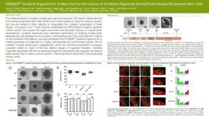 科学海报STEMdiff™ Cerebral Organoid Kit: A New Tool for the Culture of 3D Brain Organoids Derived from hPSCs


 沪公网安备31010102008431号