Balakrishnan K et al. (OCT 2006)
Blood 108 7 2392--8
Forodesine, an inhibitor of purine nucleoside phosphorylase, induces apoptosis in chronic lymphocytic leukemia cells.
Purine nucleoside phosphorylase (PNP) deficiency in humans results in T lymphocytopenia. Forodesine,a potent inhibitor of PNP,was designed based on the transition-state structure stabilized by the enzyme. Previous studies established that forodesine in the presence of deoxyguanosine (dGuo) inhibits the proliferation of T lymphocytes. A phase 1 clinical trial of forodesine in T-cell malignancies demonstrated significant antileukemic activity with an increase in intracellular dGuo triphosphate (dGTP). High accumulation of dGTP in T cells may be dependent on the levels of deoxynucleoside kinases. Because B-cell chronic lymphocytic leukemia (B-CLL) cells have high activity of deoxycytidine kinase (dCK),we hypothesized that these lymphocytes would respond to forodesine. This postulate was tested in primary lymphocytes during in vitro investigations. Lymphocytes from 12 patients with CLL were incubated with forodesine and dGuo. These CLL cells showed a wide variation in the accumulation of intracellular dGTP without any effect on other deoxynucleotides. This was associated with DNA damage-induced p53 stabilization,phosphorylation of p53 at Ser15,and activation of p21. The dGTP accumulation was related to induction of apoptosis measured by caspase activation,changes in mitochondrial membrane potential,and PARP cleavage. Based on these data,a phase 2 clinical trial of forodesine has been initiated for CLL patients.
View Publication
产品号#:
19051
19051RF
19054
19054RF
产品名:
EasySep™人T细胞富集试剂盒
RoboSep™ 人T细胞富集试剂盒含滤芯吸头
EasySep™人B细胞富集试剂盒
RoboSep™ 人B细胞富集试剂盒含滤芯吸头
Peters PJ et al. (JUL 2006)
Journal of virology 80 13 6324--32
Non-macrophage-tropic human immunodeficiency virus type 1 R5 envelopes predominate in blood, lymph nodes, and semen: implications for transmission and pathogenesis.
Human immunodeficiency virus type 1 (HIV-1) R5 isolates that predominantly use CCR5 as a coreceptor are frequently described as macrophage tropic. Here,we compare macrophage tropism conferred by HIV-1 R5 envelopes that were derived directly by PCR from patient tissue. This approach avoids potentially selective culture protocols used in virus isolation. Envelopes were amplified (i) from blood and semen of adult patients and (ii) from plasma of pediatric patients. The phenotypes of these envelopes were compared to those conferred by an extended panel of envelopes derived from brain and lymph node that we reported previously. Our results show that R5 envelopes vary by up to 1,000-fold in their capacity to confer infection of primary macrophages. Highly macrophage-tropic envelopes were predominate in brain but were infrequent in semen,blood,and lymph node samples. We also confirmed that the presence of N283 in the C2 CD4 binding site of gp120 is associated with HIV-1 envelopes from the brain but absent from macrophage-tropic envelopes amplified from blood and semen. Finally,we compared infection of macrophages,CD4(+) T cells,and peripheral blood mononuclear cells (PBMCs) conferred by macrophage-tropic and non-macrophage-tropic envelopes in the context of full-length replication competent viral clones. Non-macrophage-tropic envelopes conferred low-level infection of macrophages yet infected CD4(+) T cells and PBMCs as efficiently as highly macrophage-tropic brain envelopes. The lack of macrophage tropism for the majority of the envelopes amplified from lymph node,blood,and semen is striking and contrasts with the current consensus that R5 primary isolates are generally macrophage tropic. The extensive variation in R5 tropism reported here is likely to have an important impact on pathogenesis and on the capacity of HIV-1 to transmit.
View Publication
产品号#:
19052
19052RF
产品名:
EasySep™人CD4+ T细胞富集试剂盒
RoboSep™ 人CD4+ T细胞富集试剂盒含滤芯吸头
Zimmerman Z et al. (AUG 2005)
Biology of Blood and Marrow Transplantation 11 8 576--86
Effector cells derived from host CD8 memory T cells mediate rapid resistance against minor histocompatibility antigen-mismatched allogeneic marrow grafts without participation of perforin, Fas ligand, and the simultaneous inhibition of 3 tumor necrosis Fa
Reduced-intensity conditioning regimens for transplant recipients have heightened awareness of immunologic resistance to allogeneic bone marrow transplants (BMT). Although T cell-mediated cytotoxicity has been assumed to play a role in the resistance against donor allogeneic hematopoietic stem and progenitor cell grafts,several studies have reported relatively unimpaired resistance by recipients who lack perforin,Fas ligand (FasL),and other cytotoxic mediators. This study compared the early kinetics of T cell-mediated resistance in B6 (H2b) cytotoxically normal versus deficient recipients after transplantation with major histocompatibility complex-matched,minor histocompatibility antigen (MiHA)-mismatched allogeneic marrow grafts. Wild-type B6 or cytotoxic double-deficient perforin-/-/ gld+/+ (B6-cdd) mice were sensitized against major histocompatibility complex-matched BALB.B or C3H.SW (H2b) MiHA and transplanted with a high dose (1 ?? 107) of T cell-depleted bone marrow. CD8 T memory cells were shown to be present in recipients before BMT,and anti-CD8 monoclonal antibody infusion abolished resistance,thus demonstrating that CD8 T cells are the host effector population. Donor-committed and high proliferative potential progenitor numbers were markedly diminished by 48 hours after transplantation in both wild-type B6 and B6-cdd anti-donor MiHA-sensitized recipients. These observations indicate that the resistance pathway used in the cytotoxic deficient mice was both potent and rapidly induced - consistent with a CD8 memory T-cell response. To examine the role of Tumor necrosis factor-like weak inducer of apoptosis (TWEAK)- and TL1A-mediated cytotoxicity in this strong resistance,newly generated monoclonal antibodies specific for these ligands were administered to B6-cdd recipients sensitized to donor antigens. Recipients of syngeneic B6-gfp bone marrow exhibited significant donor colony-forming unit numbers after BMT. In contrast,low or absent colony-forming unit levels were detected in allogeneic recipients,including those that lacked perforin and FasL and that received anti-TWEAK,anti-tumor necrosis factor-related apoptosis-inducing ligand,and anti-TL1A monoclonal antibodies. These findings extend previous observations by demonstrating the existence of a rapidly effected resistance pathway mediated by memory CD8 effector T cells independent of the 2 major pathways of cytotoxicity. Together with previous findings,these results support the notion that effector cells derived from memory CD8 T-cell populations can mediate strong resistance against donor allogeneic MiHA-disparate hematopoietic engraftment by using a mechanism that is independent of the contribution of perforin,FasL,and the known death ligand receptor pathways. ?? 2005 American Society for Blood and Marrow Transplantation.
View Publication
产品号#:
03800
03801
03802
03803
03804
03805
03806
产品名:
ClonaCell™-HY杂交瘤试剂盒
ClonaCell™-HY培养基A
ClonaCell™-HY 培养基 B
ClonaCell™-HY 培养基 C
ClonaCell™-HY 培养基 D
ClonaCell™-HY 培养基 E
ClonaCell™-HY PEG
Vieillard V et al. (AUG 2005)
Proceedings of the National Academy of Sciences 102 31 10981--86
NK cytotoxicity against CD4+ T cells during HIV-1 infection: A gp41 peptide induces the expression of an NKp44 ligand
HIV infection leads to a state of chronic immune activation and progressive deterioration in immune function,manifested most recognizably by the progressive depletion of CD4+ T cells. A substantial percentage of natural killer (NK) cells from patients with HIV infection are activated and express the natural cytotoxicity receptor (NCR) NKp44. Here we show that a cellular ligand for NKp44 (NKp44L) is expressed during HIV-1 infection and is correlated with both the progression of CD4+ T cell depletion and the increase of viral load. CD4+ T cells expressing this ligand are highly sensitive to the NK lysis activity mediated by NKp44+ NK cells. The expression of NKp44L is induced by the linear motif NH2-SWSNKS-COOH of the HIV-1 envelope gp41 protein. This highly conserved motif appears critical to the sharp increase in NK lysis of CD4+ T cells from HIV-infected patients. These studies strongly suggest that induction of NKp44L plays a key role in the lysis of CD4+ T cells by activated NK cells in HIV infection and consequently provide a framework for considering how HIV-1 may use NK cell immune surveillance to trigger CD4+ T cells. Understanding this mechanism may help to develop future therapeutic strategies and vaccines against HIV-1 infection.
View Publication
产品号#:
03800
03801
03802
03803
03804
03805
03806
05150
15021
15061
产品名:
ClonaCell™-HY杂交瘤试剂盒
ClonaCell™-HY培养基A
ClonaCell™-HY 培养基 B
ClonaCell™-HY 培养基 C
ClonaCell™-HY 培养基 D
ClonaCell™-HY 培养基 E
ClonaCell™-HY PEG
MyeloCult™ H5100
RosetteSep™人T细胞富集抗体混合物
RosetteSep™人T细胞富集抗体混合物
Lalli PN et al. (SEP 2008)
Blood 112 5 1759--66
Locally produced C5a binds to T cell-expressed C5aR to enhance effector T-cell expansion by limiting antigen-induced apoptosis.
Our recent studies have shown that immune cell-produced complement provides costimulatory and survival signals to naive CD4(+) T cells. Whether these signals are similarly required during effector cell expansion and what molecular pathways link locally produced complement to T-cell survival were not clarified. To address this,we stimulated monoclonal and polyclonal T cells in vitro and in vivo with antigen-presenting cells (APCs) deficient in the complement regulatory protein,decay accelerating factor (DAF),and/or the complement component C3. We found that T-cell expansion induced by DAF-deficient APCs was augmented with diminished T-cell apoptosis,whereas T-cell expansion induced by C3(-/-) APCs was reduced because of enhanced T-cell apoptosis. These effects were traced to locally produced C5a,which through binding to T cell-expressed C5aR,enhanced expression of Bcl-2 and prevented Fas up-regulation. The results show that C5aR signal transduction in T cells is important to allow optimal T-cell expansion,as well as to maintain naive cell viability,and does so by suppressing programmed cell death.
View Publication
Le Dieu R et al. (AUG 2009)
Journal of immunological methods 348 1-2 95--100
Negative immunomagnetic selection of T cells from peripheral blood of presentation AML specimens.
To date,studies on T cells in acute myeloid leukemia (AML) have been limited to flow cytometric analysis of whole peripheral blood mononuclear cell (PBMC) specimens or functional work looking at the impact of AML myeloblasts on normal or remission T cells. This lack of information on T cells at the time of presentation with disease is due in part to the difficulty in isolating sufficiently pure T cells from these specimens for further study. Negative immunomagnetic selection has been the method of choice for isolating immune cells for functional studies due to concerns that binding antibodies to the cell surface may induce cellular activation,block ligand-receptor interactions or result in immune clearance. In order specifically to study T cells in presentation AML specimens,we set out to develop a method of isolating highly pure CD4 and CD8 T cells by negative selection from the peripheral blood (PB) of newly diagnosed AML patients. This technique,unlike T cell selection from PB from normal individuals or from patients with chronic lymphocytic leukaemia,was extremely problematic due to properties of the leukaemic myeloblasts. A successful method was eventually optimized requiring the use of a custom antibody cocktail consisting of CD33,CD34,CD123,CD11c and CD36,to deplete myeloblasts.
View Publication
产品号#:
产品名:
De Almeida DE et al. (AUG 2010)
Journal of immunology (Baltimore,Md. : 1950) 185 3 1927--34
Immune dysregulation by the rheumatoid arthritis shared epitope.
Rheumatoid arthritis (RA) is closely associated with HLA-DRB1 alleles that code a five-amino acid sequence motif in positions 70-74 of the HLA-DRbeta-chain,called the shared epitope (SE). The mechanistic basis of SE-RA association is unknown. We recently found that the SE functions as an allele-specific signal-transducing ligand that activates an NO-mediated pathway in other cells. To better understand the role of the SE in the immune system,we examined its effect on T cell polarization in mice. In CD11c(+)CD8(+) dendritic cells (DCs),the SE inhibited the enzymatic activity of indoleamine 2,3 dioxygenase,a key enzyme in immune tolerance and T cell regulation,whereas in CD11c(+)CD8(-) DCs,the ligand activated robust production of IL-6. When SE-activated DCs were cocultured with CD4(+) T cells,the differentiation of Foxp3(+) T regulatory cells was suppressed,whereas Th17 cells were expanded. The polarizing effects could be seen with SE(+) synthetic peptides,but even more so when the SE was in its natural tridimensional conformation as part of HLA-DR tetrameric proteins. In vivo administration of the SE ligand resulted in a greater abundance of Th17 cells in the draining lymph nodes and increased IL-17 production by splenocytes. Thus,we conclude that the SE acts as a potent immune-stimulatory ligand that can polarize T cell differentiation toward Th17 cells,a T cell subset that was recently implicated in the pathogenesis of autoimmune diseases,including RA.
View Publication
产品号#:
19752
19752RF
产品名:
Marks BR et al. (OCT 2009)
Nature immunology 10 10 1125--32
Thymic self-reactivity selects natural interleukin 17-producing T cells that can regulate peripheral inflammation.
Interleukin 17 (IL-17)-producing CD4(+) helper T cells (T(H)-17 cells) share a developmental relationship with Foxp3(+) regulatory T cells (T(reg) cells). Here we show that a T(H)-17 population differentiates in the thymus in a manner influenced by recognition of self antigen and by the cytokines IL-6 and transforming growth factor-beta (TGF-beta). Like previously described T(H)-17 cells,the T(H)-17 cells that developed in the thymus expressed the transcription factor RORgamma t and the IL-23 receptor. These cells also expressed alpha(4)beta(1) integrins and the chemokine receptor CCR6 and were recruited to the lung,gut and liver. In the liver,these cells secreted IL-22 in response to self antigen and mediated host protection during inflammation. Thus,T(H)-17 cells,like T(reg) cells,can be selected by self antigens in the thymus.
View Publication

 EasySep™小鼠TIL(CD45)正选试剂盒


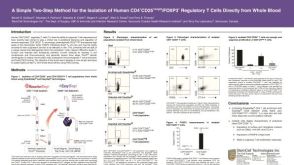 科学海报Isolation of Human CD4+CD25+Bright/Foxp3+ Regulatory T Cells Directly from Whole Blood

 沪公网安备31010102008431号