Hassanzadeh-Kiabi N et al. (NOV 2016)
Journal of immunology (Baltimore,Md. : 1950)
Autocrine Type I IFN Signaling in Dendritic Cells Stimulated with Fungal β-Glucans or Lipopolysaccharide Promotes CD8 T Cell Activation.
Type I IFNs are key mediators of immune defense against viruses and bacteria. Type I IFNs were also previously implicated in protection against fungal infection,but their roles in antifungal immunity have not been thoroughly investigated. A recent study demonstrated that bacterial and fungal β-glucans stimulate IFN-β production by dendritic cells (DCs) following detection by the Dectin-1 receptor,but the effects of β-glucan-induced type I IFNs have not been defined. We investigated whether type I IFNs regulate CD8 T cell activation by fungal β-glucan particle-stimulated DCs. We demonstrate that β-glucan-stimulated DCs induce CD8 T cell proliferation,activation marker (CD44 and CD69) expression,and production of IFN-γ,IL-2,and granzyme B. Moreover,we show that type I IFNs support robust CD8 T cell activation (proliferation and IFN-γ and granzyme B production) by β-glucan-stimulated DCs in vitro and in vivo due to autocrine effects on the DCs. Specifically,type I IFNs promote Ag presentation on MHC I molecules,CD86 and CD40 expression,and the production of IL-12 p70,IL-2,IL-6,and TNF-α by β-glucan-stimulated DCs. We also demonstrate a role for autocrine type I IFN signaling in bacterial LPS-induced DC maturation,although,in the context of LPS stimulation,this mechanism is not so critical for CD8 T cell activation (promotes IFN-γ production but not proliferation or granzyme B production). This study provides insight into the mechanisms underlying CD8 T cell activation during infection,which may be useful in the rational design of vaccines directed against pathogens and tumors.
View Publication
产品号#:
19858
19858RF
产品名:
EasySep™小鼠Naïve CD8+ T细胞分选试剂盒
RoboSep™ 小鼠Naïve CD8+ T细胞分选试剂盒
Keller G et al. (JAN 1993)
Molecular and cellular biology 13 1 473--86
Hematopoietic commitment during embryonic stem cell differentiation in culture.
We report that embryonic stem cells efficiently undergo differentiation in vitro to mesoderm and hematopoietic cells and that this in vitro system recapitulates days 6.5 to 7.5 of mouse hematopoietic development. Embryonic stem cells differentiated as embryoid bodies (EBs) develop erythroid precursors by day 4 of differentiation,and by day 6,more than 85% of EBs contain such cells. A comparative reverse transcriptase-mediated polymerase chain reaction profile of marker genes for primitive endoderm (collagen alpha IV) and mesoderm (Brachyury) indicates that both cell types are present in the developing EBs as well in normal embryos prior to the onset of hematopoiesis. GATA-1,GATA-3,and vav are expressed in both the EBs and embryos just prior to and/or during the early onset of hematopoiesis,indicating that they could play a role in the early stages of hematopoietic development both in vivo and in vitro. The initial stages of hematopoietic development within the EBs occur in the absence of added growth factors and are not significantly influenced by the addition of a broad spectrum of factors,including interleukin-3 (IL-3),IL-1,IL-6,IL-11,erythropoietin,and Kit ligand. At days 10 and 14 of differentiation,EB hematopoiesis is significantly enhanced by the addition of both Kit ligand and IL-11 to the cultures. Kinetic analysis indicates that hematopoietic precursors develop within the EBs in an ordered pattern. Precursors of the primitive erythroid lineage appear first,approximately 24 h before precursors of the macrophage and definitive erythroid lineages. Bipotential neutrophil/macrophage and multilineage precursors appear next,and precursors of the mast cell lineage develop last. The kinetics of precursor development,as well as the growth factor responsiveness of these early cells,is similar to that found in the yolk sac and early fetal liver,indicating that the onset of hematopoiesis within the EBs parallels that found in the embryo.
View Publication
产品号#:
06902
06952
00321
00322
00323
00324
00325
产品名:
挂图
Human Immune Cytokines
Infographic of key cytokines for expansion, differentiation and characterization of major immune cell types
Induction of cytotoxic T lymphocyte and antibody responses to enhanced green fluorescent protein following transplantation of transduced CD34(+) hematopoietic cells.
Genetic modification of hematopoietic stem cells often results in the expression of foreign proteins in pluripotent progenitor cells and their progeny. However,the potential for products of foreign genes introduced into hematopoietic stem cells to induce host immune responses is not well understood. Gene marking and induction of immune responses to enhanced green fluorescent protein (eGFP) were examined in rhesus macaques that underwent nonmyeloablative irradiation followed by infusions of CD34(+) bone marrow cells transduced with a retroviral vector expressing eGFP. CD34(+) cells were obtained from untreated animals or from animals treated with recombinant human granulocyte colony-stimulating factor (G-CSF) alone or G-CSF and recombinant human stem cell factor. Levels of eGFP-expressing cells detected by flow cytometry peaked at 0.1% to 0.5% of all leukocytes 1 to 4 weeks after transplantation. Proviral DNA was detected in 0% to 17% of bone marrow--derived colony-forming units at periods of 5 to 18 weeks after transplantation. However,5 of 6 animals studied demonstrated a vigorous eGFP-specific cytotoxic T lymphocyte (CTL) response that was associated with a loss of genetically modified cells in peripheral blood,as demonstrated by both flow cytometry and polymerase chain reaction. The eGFP-specific CTL responses were MHC-restricted,mediated by CD8(+) lymphocytes,and directed against multiple epitopes. eGFP-specific CTLs were able to efficiently lyse autologous CD34(+) cells expressing eGFP. Antibody responses to eGFP were detected in 3 of 6 animals. These data document the potential for foreign proteins expressed in CD34(+) hematopoietic cells and their progeny to induce antibody and CTL responses in the setting of a clinically applicable transplantation protocol. (Blood. 2001;97:1951-1959)
View Publication

 EasySep™小鼠TIL(CD45)正选试剂盒


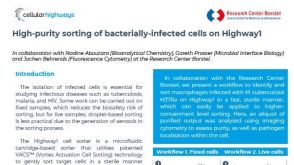
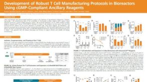 科学海报Development of Robust T Cell Manufacturing Protocols in Bioreactors Using cGMP-Compliant Ancillary Reagents
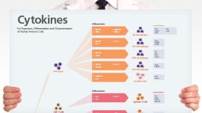 挂图Human Immune Cytokines Infographic of key cytokines for expansion, differentiation and characterization of major immune cell types
 技术公告Achieve Scalable, High-Quality Nucleic Acid Extractions with the EasySep™ Total Nucleic Acid Extraction Kit


 沪公网安备31010102008431号