Carroll VA et al. (OCT 2016)
Proceedings of the National Academy of Sciences of the United States of America
Expression of HIV-1 matrix protein p17 and association with B-cell lymphoma in HIV-1 transgenic mice.
HIV-1 infection is associated with increased risk for B-cell lymphomas. How HIV infection promotes the development of lymphoma is unclear,but it may involve chronic B-cell activation,inflammation,and/or impaired immunity,possibly leading to a loss of control of oncogenic viruses and reduced tumor immunosurveillance. We hypothesized that HIV structural proteins may contribute to lymphomagenesis directly,because they can persist long term in lymph nodes in the absence of viral replication. The HIV-1 transgenic mouse Tg26 carries a noninfectious HIV-1 provirus lacking part of the gag-pol region,thus constituting a model for studying the effects of viral products in pathogenesis. Approximately 15% of Tg26 mice spontaneously develop leukemia/lymphoma. We investigated which viral proteins are associated with the development of leukemia/lymphoma in the Tg26 mouse model,and performed microarray analysis on RNA from spleen and lymph nodes to identify potential mechanisms of lymphomagenesis. Of the viral proteins examined,only expression of HIV-1 matrix protein p17 was associated with leukemia/lymphoma development and was highly expressed in bone marrow before disease. The tumor cells resembled pro-B cells,and were CD19(+)IgM(-)IgD(-)CD93(+)CD43(+)CD21(-)CD23(-)VpreB(+)CXCR4(+) Consistent with the pro-B-cell stage of B-cell development,microarray analysis revealed enrichment of transcripts,including Rag1,Rag2,CD93,Vpreb1,Vpreb3,and Igll1 We confirmed RAG1 expression in Tg26 tumors,and hypothesized that HIV-1 matrix protein p17 may directly induce RAG1 in B cells. Stimulation of human activated B cells with p17 enhanced RAG1 expression in three of seven donors,suggesting that intracellular signaling by p17 may lead to genomic instability and transformation.
View Publication
产品号#:
19054
19054RF
17963
17963RF
产品名:
EasySep™人B细胞富集试剂盒
RoboSep™ 人B细胞富集试剂盒含滤芯吸头
EasySep™人B细胞富集试剂盒II(不去除CD43)
RoboSep™ 人B细胞富集试剂盒II(不去除CD43)
Chalmers SA et al. (MAY 2016)
Scientific Reports 6 26164
Therapeutic Blockade of Immune Complex-Mediated Glomerulonephritis by Highly Selective Inhibition of Bruton's Tyrosine Kinase.
Lupus nephritis (LN) is a potentially dangerous end organ pathology that affects upwards of 60% of lupus patients. Bruton's tyrosine kinase (BTK) is important for B cell development,Fc receptor signaling,and macrophage polarization. In this study,we investigated the effects of a novel,highly selective and potent BTK inhibitor,BI-BTK-1,in an inducible model of LN in which mice receive nephrotoxic serum (NTS) containing anti-glomerular antibodies. Mice were treated once daily with vehicle alone or BI-BTK-1,either prophylactically or therapeutically. When compared with control treated mice,NTS-challenged mice treated prophylactically with BI-BTK-1 exhibited significantly attenuated kidney disease,which was dose dependent. BI-BTK-1 treatment resulted in decreased infiltrating IBA-1+ cells,as well as C3 deposition within the kidney. RT-PCR on whole kidney RNA and serum profiling indicated that BTK inhibition significantly decreased levels of LN-relevant inflammatory cytokines and chemokines. Renal RNA expression profiling by RNA-seq revealed that BI-BTK-1 dramatically modulated pathways related to inflammation and glomerular injury. Importantly,when administered therapeutically,BI-BTK-1 reversed established proteinuria and improved renal histopathology. Our results highlight the important role for BTK in the pathogenesis of immune complex-mediated nephritis,and BTK inhibition as a promising therapeutic target for LN.
View Publication
产品号#:
19359
19359RF
19054
19054RF
100-0697
产品名:
EasySep™人单核细胞分选试剂盒
RoboSep™ 人单核细胞分选试剂盒
EasySep™人B细胞富集试剂盒
RoboSep™ 人B细胞富集试剂盒含滤芯吸头
EasySep™人单核细胞分选试剂盒
Ciurea SO et al. (AUG 2007)
Blood 110 3 986--93
Pivotal contributions of megakaryocytes to the biology of idiopathic myelofibrosis.
In order to investigate the biologic processes underlying and resulting from the megakaryocytic hyperplasia that characterizes idiopathic myelofibrosis (IMF),peripheral blood CD34+ cells isolated from patients with IMF,polycythemia vera (PV),and G-CSF-mobilized healthy volunteers were cultured in the presence of stem cell factor and thrombopoietin. IMF CD34+ cells generated 24-fold greater numbers of megakaryocytes (MKs) than normal CD34+ cells. IMF MKs were also shown to have a delayed pattern of apoptosis and to overexpress the antiapoptotic protein bcl-xL. MK hyperplasia in IMF is,therefore,likely a consequence of both the increased ability of IMF progenitor cells to generate MKs and a decreased rate of MK apoptosis. Media conditioned (CM) by CD61+ cells generated in vitro from CD34+ cells were then assayed for the levels of growth factors and proteases. Higher levels of transforming growth factor-beta (TGF-beta) and active matrix metalloproteinase-9 (MMP9) were observed in media conditioned with IMF CD61+ cells than normal or PV CD61+ cells. Both normal and IMF CD61+ cells produced similar levels of VEGF. MK-derived TGF-B and MMP-9,therefore,likely contribute to the development of many pathological epiphenomena associated with IMF.
View Publication
产品号#:
09600
09650
产品名:
StemSpan™ SFEM
StemSpan™ SFEM
Walker WE et al. (OCT 2006)
Journal of immunology (Baltimore,Md. : 1950) 177 8 5307--16
Absence of innate MyD88 signaling promotes inducible allograft acceptance.
Prior experimental strategies to induce transplantation tolerance have focused largely on modifying adaptive immunity. However,less is known concerning the role of innate immune signaling in the induction of transplantation tolerance. Using a highly immunogenic murine skin transplant model that resists transplantation tolerance induction when innate immunity is preserved,we show that absence of MyD88,a key innate Toll like receptor signal adaptor,abrogates this resistance and facilitates inducible allograft acceptance. In our model,absence of MyD88 impairs inflammatory dendritic cell responses that reduce T cell activation. This effect increases T cell susceptibility to suppression mediated by CD4+ CD25+ regulatory T cells. Therefore,this study provides evidence that absence of MyD88 promotes inducible allograft acceptance and implies that inhibiting innate immunity may be a potential,clinically relevant strategy to facilitate transplantation tolerance.
View Publication
产品号#:
18758
18758RF
18768
18768RF
19752
19752RF
19753
19753RF
产品名:
Irish JM et al. (AUG 2006)
Journal of immunology (Baltimore,Md. : 1950) 177 3 1581--9
Kinetics of B cell receptor signaling in human B cell subsets mapped by phosphospecific flow cytometry.
Differences in BCR signaling may govern outcomes as diverse as proliferation and cell death. We profiled BCR signaling kinetics in subsets of primary human B cells using flow cytometry. In the predominant population expressing IgM,BCR cross-linking led to a quick burst of Syk,ERK1/2,and p38 signaling. In contrast,IgG B cells sustained higher per-cell ERK1/2 phosphorylation over time. This dichotomy suggested a mechanism for dampening signals transmitted by IgM. Regulatory phosphatase activity in IgM B cells was BCR-mediated and initiated more slowly than kinase activity. This BCR-mediated phosphatase activity was sensitive to inhibition by H(2)O(2) and required to attenuate IgM BCR signaling. These results provide the first kinetic maps of BCR signaling in primary human B cell subsets and enable new studies of signaling in B cell disorders,such as autoimmunity and cancer.
View Publication
Seo J-H et al. (SEP 2010)
Cancer research 70 18 7325--35
A specific need for CRKL in p210BCR-ABL-induced transformation of mouse hematopoietic progenitors.
CRKL (CRK-like) is an adapter protein predominantly phosphorylated in cells that express the tyrosine kinase p210(BCR-ABL),the fusion product of a (9;22) chromosomal translocation causative for chronic myeloid leukemia. It has been unclear,however,whether CRKL plays a functional role in p210(BCR-ABL) transformation. Here,we show that CRKL is required for p210(BCR-ABL) to support interleukin-3-independent growth of myeloid progenitor cells and long-term outgrowth of B-lymphoid cells from fetal liver-derived hematopoietic progenitor cells. Furthermore,a synthetic phosphotyrosyl peptide that binds to the CRKL SH2 domain with high affinity blocks association of endogenous CRKL with the p210(BCR-ABL) complex and reduces c-MYC levels in K562 human leukemic cells as well as in mouse hematopoietic cells transformed by p210(BCR-ABL) or the imatinib-resistant mutant T315I. These results indicate that the function of CRKL as an adapter protein is essential for p210(BCR-ABL)-induced transformation.
View Publication
Agosti V et al. (MAR 2004)
The Journal of experimental medicine 199 6 867--78
Critical role for Kit-mediated Src kinase but not PI 3-kinase signaling in pro T and pro B cell development.
The Kit receptor functions in hematopoiesis,lymphocyte development,gastrointestinal tract motility,melanogenesis,and gametogenesis. To investigate the roles of different Kit signaling pathways in vivo,we have generated knock-in mice in which docking sites for PI 3-kinase (KitY719) or Src kinase (KitY567) have been mutated. Whereas steady-state hematopoiesis is normal in KitY719F/Y719F and KitY567F/Y567F mice,lymphopoiesis is affected differentially. The KitY567F mutation,but not the KitY719F mutation,blocks pro T cell and pro B cell development in an age-dependent manner. Thus,the Src family kinase,but not the PI 3-kinase docking site in Kit,mediates a critical signal for lymphocyte development. In agreement with these results,treatment of normal mice with the Kit tyrosine kinase inhibitor imatinib (Gleevec) leads to deficits in pro T and pro B cell development,similar to those seen in KitY567F/Y567F and KitW/W mice. The two mutations do not affect embryonic gametogenesis but the KitY719F mutation blocks spermatogenesis at the spermatogonial stages and in contrast the KitY567F mutation does not affect this process. Therefore,Kit-mediated PI 3-kinase signaling and Src kinase family signaling is highly specific for different cellular contexts in vivo.
View Publication

 EasySep™小鼠TIL(CD45)正选试剂盒


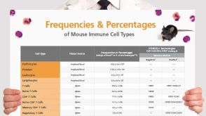 挂图Frequencies and Percentages of Mouse Immune Cell Types List of the frequencies of over 25 immune cell types in C57BL/6 mice
 沪公网安备31010102008431号