Li MMH et al. (NOV 2016)
The Journal of experimental medicine
Interferon regulatory factor 2 protects mice from lethal viral neuroinvasion.
The host responds to virus infection by activating type I interferon (IFN) signaling leading to expression of IFN-stimulated genes (ISGs). Dysregulation of the IFN response results in inflammatory diseases and chronic infections. In this study,we demonstrate that IFN regulatory factor 2 (IRF2),an ISG and a negative regulator of IFN signaling,influences alphavirus neuroinvasion and pathogenesis. A Sindbis virus strain that in wild-type (WT) mice only causes disease when injected into the brain leads to lethal encephalitis in Irf2(-/-) mice after peripheral inoculation. Irf2(-/-) mice fail to control virus replication and recruit immune infiltrates into the brain. Reduced B cells and virus-specific IgG are observed in the Irf2(-/-) mouse brains despite the presence of peripheral neutralizing antibodies,suggesting a defect in B cell trafficking to the central nervous system (CNS). B cell-deficient μMT mice are significantly more susceptible to viral infection,yet WT B cells and serum are unable to rescue the Irf2(-/-) mice. Collectively,our data demonstrate that proper localization of B cells and local production of antibodies in the CNS are required for protection. The work advances our understanding of host mechanisms that affect viral neuroinvasion and their contribution to immunity against CNS infections.
View Publication
产品号#:
19854
19854RF
产品名:
EasySep™小鼠B细胞分选试剂盒
RoboSep™ 小鼠B细胞分选试剂盒
Li H et al. (AUG 2010)
Blood 116 7 1060--9
Repression of Id2 expression by Gfi-1 is required for B-cell and myeloid development.
The development of mature blood cells from hematopoietic stem cells requires coordinated activities of transcriptional networks. Transcriptional repressor growth factor independence 1 (Gfi-1) is required for the development of B cells,T cells,neutrophils,and for the maintenance of hematopoietic stem cell function. However,the mechanisms by which Gfi-1 regulates hematopoiesis and how Gfi-1 integrates into transcriptional networks remain unclear. Here,we provide evidence that Id2 is a transcriptional target of Gfi-1,and repression of Id2 by Gfi-1 is required for B-cell and myeloid development. Gfi-1 binds to 3 conserved regions in the Id2 promoter and represses Id2 promoter activity in transient reporter assays. Increased Id2 expression was observed in multipotent progenitors,myeloid progenitors,T-cell progenitors,and B-cell progenitors in Gfi-1(-/-) mice. Knockdown of Id2 expression or heterozygosity at the Id2 locus partially rescues the B-cell and myeloid development but not the T-cell development in Gfi-1(-/-) mice. These studies demonstrate a role of Id2 in mediating Gfi-1 functions in B-cell and myeloid development and provide a direct link between Gfi-1 and the B-cell transcriptional network by its ability to repress Id2 expression.
View Publication
产品号#:
03234
产品名:
MethoCult™ M3234
Inoue S et al. (AUG 2006)
Cancer research 66 15 7741--7
Inhibitory effects of B cells on antitumor immunity.
B-cell functions in antitumor immunity are not well understood. In this study,we evaluated the role of B cells in the development of antitumor immunity using Friend murine leukemia virus gag-expressing mouse EL-4 (EL-4 gag),D5 mouse melanoma,or MCA304 mouse sarcoma cells. To screen tumors for susceptibility to B-cell-deficient immune environments,spleen cells from naive C57BL/6 [wild-type (WT)] and B-cell knockout (BKO) mice were cultured with irradiated tumor cells in vitro. When cells were stimulated with EL-4 gag or D5 (but not MCA304 tumors),IFN-gamma production from CD8 T cells and natural killer cells was markedly decreased in WT compared with BKO cultures. IFN-gamma production was correlated with CD40 ligand expression on the tumor and inversely with interleukin-10 (IL-10) production by B cells. Sorted WT B cells produced more IL-10 than CD40 knockout (CD40KO) B cells when cocultured with EL-4 gag or D5 (but not MCA304). IFN-gamma production by BKO cells was reduced by the addition of sorted naive WT B cells (partially by CD40KO B cells) or recombinant mouse IL-10. In vivo tumor progression mirrored in vitro studies in that WT mice were unable to control tumor growth whereas EL-4 gag and D5 tumors (but not MCA304) were eliminated in BKO mice. Robust in vivo antitumor CTLs developed only in BKO tumor-challenged mice. Our studies provide the first mechanistic basis for the concept that B-cell depletion could therapeutically enhance antitumor immune responses to certain tumors by decreasing IL-10 production from B cells.
View Publication
产品号#:
18754
18754RF
产品名:
Giassi LJ et al. (AUG 2008)
Experimental biology and medicine (Maywood,N.J.) 233 8 997--1012
Expanded CD34+ human umbilical cord blood cells generate multiple lymphohematopoietic lineages in NOD-scid IL2rgamma(null) mice.
Umbilical cord blood (UCB) is increasingly being used for human hematopoietic stem cell (HSC) transplantation in children but often requires pooling multiple cords to obtain sufficient numbers for transplantation in adults. To overcome this limitation,we have used an ex vivo two-week culture system to expand the number of hematopoietic CD34(+) cells in cord blood. To assess the in vivo function of these expanded CD34(+) cells,cultured human UCB containing 1 x 10(6) CD34(+) cells were transplanted into conditioned NOD-scid IL2rgamma(null) mice. The expanded CD34(+) cells displayed short- and long-term repopulating cell activity. The cultured human cells differentiated into myeloid,B-lymphoid,and erythroid lineages,but not T lymphocytes. Administration of human recombinant TNFalpha to recipient mice immediately prior to transplantation promoted human thymocyte and T-cell development. These T cells proliferated vigorously in response to TCR cross-linking by anti-CD3 antibody. Engrafted TNFalpha-treated mice generated antibodies in response to T-dependent and T-independent immunization,which was enhanced when mice were co-treated with the B cell cytokine BLyS. Ex vivo expanded CD34(+) human UCB cells have the capacity to generate multiple hematopoietic lineages and a functional human immune system upon transplantation into TNFalpha-treated NOD-scid IL2rgamma(null) mice.
View Publication
产品号#:
09600
09650
09850
产品名:
StemSpan™ SFEM
StemSpan™ SFEM
Doreau A et al. (JUL 2009)
Nature immunology 10 7 778--85
Interleukin 17 acts in synergy with B cell-activating factor to influence B cell biology and the pathophysiology of systemic lupus erythematosus.
Studies have suggested involvement of interleukin 17 (IL-17) in autoimmune diseases,although its effect on B cell biology has not been clearly established. Here we demonstrate that IL-17 alone or in combination with B cell-activating factor controlled the survival and proliferation of human B cells and their differentiation into immunoglobulin-secreting cells. This effect was mediated mainly through the nuclear factor-kappaB-regulated transcription factor Twist-1. In support of the relevance of our observations and the potential involvement of IL-17 in B cell biology,we found that the serum of patients with systemic lupus erythematosus had higher concentrations of IL-17 than did the serum of healthy people and that IL-17 abundance correlated with the disease severity of systemic lupus erythematosus.
View Publication
产品号#:
18054
18054RF
产品名:
Irish JM et al. (NOV 2006)
Blood 108 9 3135--42
Altered B-cell receptor signaling kinetics distinguish human follicular lymphoma B cells from tumor-infiltrating nonmalignant B cells.
The B-cell receptor (BCR) transmits life and death signals throughout B-cell development,and altered BCR signaling may be required for survival of B-lymphoma cells. We used single-cell signaling profiles to compare follicular lymphoma (FL) B cells and nonmalignant host B cells within individual patient biopsies and identified BCR-mediated signaling events specific to lymphoma B cells. Expression of CD20,Bcl-2,and BCR light chain isotype (kappa or lambda) distinguished FL tumor B-cell and nontumor host B-cell subsets within FL patient biopsies. BCR-mediated signaling via phosphorylation of Btk,Syk,Erk1/2,and p38 occurred more rapidly in tumor B cells from FL samples than in infiltrating nontumor B cells,achieved greater levels of per-cell signaling,and sustained this level of signaling for hours longer than nontumor B cells. The timing and magnitude of BCR-mediated signaling in nontumor B cells within an FL sample instead resembled that observed in mature B cells from the peripheral blood of healthy subjects. BCR signaling pathways that are potentiated specifically in lymphoma cells should provide new targets for therapeutic attention.
View Publication
产品号#:
09850
产品名:
Mihalcik SA et al. (JUL 2010)
Journal of immunology (Baltimore,Md. : 1950) 185 2 1045--54
The structure of the TNFRSF13C promoter enables differential expression of BAFF-R during B cell ontogeny and terminal differentiation.
The B cell-activating factor of the TNF family receptor (BAFF-R),encoded by the TNFRSF13C gene,is critically important for transitional B cell survival to maturity. Thus,ligation of BAFF-R by BAFF delivers a potent survival signal. Reports implicating the BAFF/BAFF-R signaling axis in the pathogenesis of autoimmune human diseases and B lineage malignancies have largely prompted studies focusing on BAFF expression; however,there is an equally critical need to better understand BAFF-R expression. Initial BAFF-R expression,although characterized in murine B cells,has not yet been reported in human B lymphopoiesis. In this study,we first demonstrate that BAFF-R expression is absent from early precursors and is acquired by bone marrow B cells newly expressing the BCR. We next focused on identifying the specific genomic region that controls BAFF-R expression in mature B cells (i.e.,the TNFRSF13C promoter). To accomplish this,we used in silico tools examining interspecies genomic conservation in conjunction with reporter constructs transfected into malignant B and plasma cell lines. DNase protection assays using nuclear extracts from BAFF-R-expressing cells suggested potential regulatory sites,which allowed the generation of EMSA probes that bound NFs specific to BAFF-R-expressing cells. With a more stringent analysis of interspecies homology,these assays identified a site at which a single nucleotide substitution could distinctly impact promoter activity. Finally,chromatin immunoprecipitation assays revealed the in vivo binding of the specific transcription factor c-Rel to the most proximal genomic region,and c-Rel small interfering RNA transfections in BAFF-R-expressing lines demonstrated a coincident knockdown of both c-Rel and BAFF-R mRNA.
View Publication

 EasySep™小鼠TIL(CD45)正选试剂盒


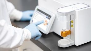 实验方案Optimizing Delivery Efficiency with Fluorescent Dextran Using the CellPore™ Transfection System

 沪公网安备31010102008431号