Nguyen CQ et al. (JUL 2007)
Journal of immunology (Baltimore,Md. : 1950) 179 1 382--90
IL-4-STAT6 signal transduction-dependent induction of the clinical phase of Sjögren's syndrome-like disease of the nonobese diabetic mouse.
NOD.B10-H2(b) and NOD/LtJ mice manifest,respectively,many features of primary and secondary Sjögren's syndrome (SjS),an autoimmune disease affecting primarily the salivary and lacrimal glands leading to xerostomia (dry mouth) and xerophthalmia (dry eyes). B lymphocytes play a central role in the onset of SjS with clinical manifestations dependent on the appearance of autoantibodies reactive to multiple components of acinar cells. Previous studies with NOD.IL4(-/-) and NOD.B10-H2(b).IL4(-/-) mice suggest that the Th2 cytokine,IL-4,plays a vital role in the development and onset of SjS-like disease in the NOD mouse model. To investigate the molecular mechanisms by which IL-4 controls SjS development,a Stat6 gene knockout mouse,NOD.B10-H2(b).C-Stat6(-/-),was constructed and its disease profile was defined and compared with that of NOD.B10-H2(b).C-Stat6(+/+) mice. As the NOD.B10-H2(b).C-Stat6(-/-) mice aged from 4 to 24 wk,they exhibited leukocyte infiltration of the exocrine glands,produced anti-nuclear autoantibodies,and showed loss and gain of saliva-associated proteolytic enzymes,similar to NOD.B10-H2(b).C-Stat6(+/+) mice. In contrast,NOD.B10-H2(b).C-Stat6(-/-) mice failed to develop glandular dysfunction,maintaining normal saliva flow rates. NOD.B10-H2(b).C-Stat6(-/-) mice were found to lack IgG1 isotype-specific anti-muscarinic acetylcholine type-3 receptor autoantibodies. Furthermore,the IgG fractions from NOD.B10-H2(b).C-Stat6(-/-) sera were unable to induce glandular dysfunction when injected into naive recipient C57BL/6 mice. NOD.B10-H2(b).C-Stat6(-/-) mice,like NOD.B10-H2(b).IL4(-/-) mice,are unable to synthesize IgG1 Abs,an observation that correlates with an inability to develop end-stage clinical SjS-like disease. These data imply a requirement for the IL-4/STAT6-pathway for onset of the clinical phase of SjS-like disease in the NOD mouse model.
View Publication
产品号#:
18754
18754RF
产品名:
Smith KS et al. (NOV 2002)
Molecular and cellular biology 22 21 7678--87
Transformation of bone marrow B-cell progenitors by E2a-Hlf requires coexpression of Bcl-2.
The chimeric transcription factor E2a-Hlf is an oncoprotein associated with a subset of acute lymphoblastic leukemias of early B-lineage derivation. We employed a retroviral transduction-transplantation approach to evaluate the oncogenic effects of E2a-Hlf on murine B-cell progenitors harvested from adult bone marrow. Expression of E2a-Hlf induced short-lived clusters of primary hematopoietic cells but no long-term growth on preformed bone marrow stromal cell layers comprised of the AC6.21 cell line. Coexpression with Bcl-2,however,resulted in the sustained self-renewal of early preB-I cells that required stromal and interleukin-7 (IL-7) support for growth in vitro. Immortalized cells were unable to induce leukemias after transplantation into nonirradiated syngeneic hosts,unlike the leukemic properties and cytokine independence of preB-I cells transformed by p190(Bcr-Abl) under identical in vitro conditions. However,bone marrow cells expressing E2a-Hlf in combination with Bcl-2,but not E2a-Hlf alone,induced leukemias in irradiated recipients with long latencies,demonstrating both a requirement for suppression of apoptosis and the need for further secondary mutations in leukemia pathogenesis. Coexpression of IL-7 substituted for Bcl-2 to induce the in vitro growth of pre-B cells expressing E2a-Hlf,but leukemic conversion required additional abrogation of undefined stromal requirements and was associated with alterations in the Arf/Mdm2/p53 pathway. Thus,E2a-Hlf enhances the self-renewal of bone marrow B-cell progenitors without inciting a p53 tumor surveillance response or abrogating stromal and cytokine requirements for growth,which are nevertheless abrogated during progression to a leukemogenic phenotype.
View Publication
产品号#:
03134
产品名:
MethoCult™ M3134
Yates F et al. (DEC 2002)
Blood 100 12 3942--9
Gene therapy of RAG-2-/- mice: sustained correction of the immunodeficiency.
Patients with mutations of either RAG-1 or RAG-2 genes suffer from severe combined immunodeficiency (SCID) characterized by the lack of T and B lymphocytes. The only curative treatment today consists of hematopoietic stem cell (HSC) transplantation,which is only partially successful in the absence of an HLA genoidentical donor,thus justifying research to find an alternative therapeutic approach. To this end,RAG-2-deficient mice were used to test whether retrovirally mediated ex vivo gene transfer into HSCs could provide long-term correction of the immunologic deficiency. Murine RAG-2-/-Sca-1(+) selected bone marrow cells were transduced with a modified Moloney leukemia virus (MLV)-based MND (myeloproliferative sarcoma virus enhancer,negative control region deleted,dl587rev primer-binding site substituted) retroviral vector containing the RAG-2 cDNA and transplanted into RAG-2-/- sublethally irradiated mice (3Gy). Two months later,T- and B-cell development was achieved in all mice. Diverse repertoire of T cells as well as proliferative capacity in the presence of mitogens,allogeneic cells,and keyhole limpet hemocyanin (KLH) were shown. B-cell function as shown by serum Ig levels and antibody response to a challenge by KLH also developed. Lymphoid subsets and function were shown to be stable over a one-year period without evidence of any detectable toxicity. Noteworthy,a selective advantage for transduced lymphoid cells was evidenced by comparative provirus quantification in lymphoid and myeloid lineages. Altogether,this study demonstrates the efficiency of ex vivo RAG-2 gene transfer in HSCs to correct the immune deficiency of RAG-2-/- mice,constituting a significant step toward clinical application.
View Publication
产品号#:
09600
09650
产品名:
StemSpan™ SFEM
StemSpan™ SFEM
Glodek AM et al. (FEB 2003)
The Journal of experimental medicine 197 4 461--73
Sustained activation of cell adhesion is a differentially regulated process in B lymphopoiesis.
It is largely unknown how hematopoietic progenitors are positioned within specialized niches of the bone marrow microenvironment during development. Chemokines such as CXCL12,previously called stromal cell-derived factor 1,are known to activate cell integrins of circulating leukocytes resulting in transient adhesion before extravasation into tissues. However,this short-term effect does not explain the mechanism by which progenitor cells are retained for prolonged periods in the bone marrow. Here we show that in human bone marrow CXCL12 triggers a sustained adhesion response specifically in progenitor (pro- and pre-) B cells. This sustained adhesion diminishes during B cell maturation in the bone marrow and,strikingly,is absent in circulating mature B cells,which exhibit only transient CXCL12-induced adhesion. The duration of adhesion is tightly correlated with CXCL12-induced activation of focal adhesion kinase (FAK),a known molecule involved in integrin-mediated signaling. Sustained adhesion of progenitor B cells is associated with prolonged FAK activation,whereas transient adhesion in circulating B cells is associated with short-lived FAK activation. Moreover,sustained and transient adhesion responses are differentially affected by pharmacological inhibitors of protein kinase C and phosphatidylinositol 3-kinase. These results provide a developmental cell stage-specific mechanism by which chemokines orchestrate hematopoiesis through sustained rather than transient activation of adhesion and cell survival pathways.
View Publication
产品号#:
产品名:
Iwasaki-Arai J et al. (MAY 2003)
The Journal of experimental medicine 197 10 1311--22
Enforced granulocyte/macrophage colony-stimulating factor signals do not support lymphopoiesis, but instruct lymphoid to myelomonocytic lineage conversion.
We evaluated the effects of ectopic granulocyte/macrophage colony-stimulating factor (GM-CSF) signals on hematopoietic commitment and differentiation. Lineage-restricted progenitors purified from mice with the ubiquitous transgenic human GM-CSF receptor (hGM-CSFR) were used for the analysis. In cultures with hGM-CSF alone,hGM-CSFR-expressing (hGM-CSFR+) granulocyte/monocyte progenitors (GMPs) and megakaryocyte/erythrocyte progenitors (MEPs) exclusively gave rise to granulocyte/monocyte (GM) and megakaryocyte/erythroid (MegE) colonies,respectively,providing formal proof that GM-CSF signals support the GM and MegE lineage differentiation without affecting the physiological myeloid fate. hGM-CSFR transgenic mice were crossed with mice deficient in interleukin (IL)-7,an essential cytokine for T and B cell development. Administration of hGM-CSF in these mice could not restore T or B lymphopoiesis,indicating that enforced GM-CSF signals cannot substitute for IL-7 to promote lymphopoiesis. Strikingly,textgreater50% hGM-CSFR+ common lymphoid progenitors (CLPs) and textgreater20% hGM-CSFR+ pro-T cells gave rise to granulocyte,monocyte,and/or myeloid dendritic cells,but not MegE lineage cells in the presence of hGM-CSF. Injection of hGM-CSF into mice transplanted with hGM-CSFR+ CLPs blocked their lymphoid differentiation,but induced development of GM cells in vivo. Thus,hGM-CSF transduces permissive signals for myeloerythroid differentiation,whereas it transmits potent instructive signals for the GM differentiation to CLPs and early T cell progenitors. These data suggest that a majority of CLPs and a fraction of pro-T cells possess plasticity for myelomonocytic differentiation that can be activated by ectopic GM-CSF signals,supporting the hypothesis that the down-regulation of GM-CSFR is a critical event in producing cells with a lymphoid-restricted lineage potential.
View Publication
产品号#:
04100
产品名:
MethoCult™ H4100
Coletta PL et al. (FEB 2004)
Blood 103 3 1050--8
Lymphodepletion in the ApcMin/+ mouse model of intestinal tumorigenesis.
Germ line mutations in the Adenomatous polyposis coli tumor suppressor gene cause a hereditary form of intestinal tumorigenesis in both mice and man. Here we show that in Apc(Min/+) mice,which carry a heterozygous germ line mutation at codon 850 of Apc,there is progressive loss of immature and mature thymocytes from approximately 80 days of age with complete regression of the thymus by 120 days. In addition,Apc(Min/+) mice show parallel depletion of splenic natural killer (NK) cells,immature B cells,and B progenitor cells in bone marrow due to complete loss of interleukin 7 (IL-7)-dependent B-cell progenitors. Using bone marrow transplantation experiments into wild-type recipients,we have shown that the capacity of transplanted Apc(Min/+) bone marrow cells for T- and B-cell development appears normal. In contrast,although the Apc(Min/+) bone marrow microenvironment supported short-term reconstitution with wild-type bone marrow,Apc(Min/+) animals that received transplants subsequently underwent lymphodepletion. Fibroblast colony-forming unit (CFU-F) colony assays revealed a significant reduction in colony-forming mesenchymal progenitor cells in the bone marrow of Apc(Min/+) mice compared with wild-type animals prior to the onset of lymphodepletion. This suggests that an altered bone marrow microenvironment may account for the selective lymphocyte depletion observed in this model of familial adenomatous polyposis.
View Publication
产品号#:
03630
03434
03444
05501
05502
产品名:
MethoCult™ M3630
MethoCult™ GF M3434
MethoCult™ GF M3434
Morrow M et al. (MAY 2004)
Blood 103 10 3890--6
TEL-AML1 promotes development of specific hematopoietic lineages consistent with preleukemic activity.
The t(12;21)(p13;q22) translocation is the most common chromosomal abnormality yet identified in any pediatric leukemia and gives rise to the TEL-AML1 fusion product. To investigate the effects of TEL-AML1 on hematopoiesis,fetal liver hematopoietic progenitor cells (HPCs) were transduced with retroviral vectors expressing this fusion protein. We show that TEL-AML1 dramatically alters differentiation of HPCs in vitro,preferentially promoting B-lymphocyte development,enhancing self-renewal of B-cell precursors,and leading to the establishment of long-term growth factor-dependent pre-B-cell lines. However,it had no effect on myeloid development in vitro. Further experiments were performed to determine whether TEL-AML1 also demonstrates lineage-specific activity in vivo. TEL-AML1-expressing HPCs displayed a competitive advantage in reconstituting both B-cell and myeloid lineages in vivo but had no effect on reconstitution of the T-cell lineage. Despite promoting these alterations in hematopoiesis,TEL-AML1 did not induce leukemia in transplanted mice. Our study provides a unique insight into the role of TEL-AML1 in leukemia predisposition and a potential model to study the mechanism of leukemogenesis associated with this fusion.
View Publication

 EasySep™小鼠TIL(CD45)正选试剂盒


 技术公告Achieve Scalable, High-Quality Nucleic Acid Extractions with the EasySep™ Total Nucleic Acid Extraction Kit
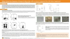 科学海报Robust Serum-Free Expansion of Human B Cells In Vitro with an Animal Component-Free Cell Culture Supplement


 沪公网安备31010102008431号