Application of the pMHC Array to Characterise Tumour Antigen Specific T Cell Populations in Leukaemia Patients at Disease Diagnosis.
Immunotherapy treatments for cancer are becoming increasingly successful,however to further improve our understanding of the T-cell recognition involved in effective responses and to encourage moves towards the development of personalised treatments for leukaemia immunotherapy,precise antigenic targets in individual patients have been identified. Cellular arrays using peptide-MHC (pMHC) tetramers allow the simultaneous detection of different antigen specific T-cell populations naturally circulating in patients and normal donors. We have developed the pMHC array to detect CD8+ T-cell populations in leukaemia patients that recognise epitopes within viral antigens (cytomegalovirus (CMV) and influenza (Flu)) and leukaemia antigens (including Per Arnt Sim domain 1 (PASD1),MelanA,Wilms' Tumour (WT1) and tyrosinase). We show that the pMHC array is at least as sensitive as flow cytometry and has the potential to rapidly identify more than 40 specific T-cell populations in a small sample of T-cells (0.8-1.4 x 10(6)). Fourteen of the twenty-six acute myeloid leukaemia (AML) patients analysed had T cells that recognised tumour antigen epitopes,and eight of these recognised PASD1 epitopes. Other tumour epitopes recognised were MelanA (n = 3),tyrosinase (n = 3) and WT1(126-134) (n = 1). One of the seven acute lymphocytic leukaemia (ALL) patients analysed had T cells that recognised the MUC1(950-958) epitope. In the future the pMHC array may be used provide point of care T-cell analyses,predict patient response to conventional therapy and direct personalised immunotherapy for patients.
View Publication
产品号#:
19053
19053RF
产品名:
EasySep™人CD8+ T细胞富集试剂盒
RoboSep™ 人CD8+ T细胞富集试剂盒含滤芯吸头
Gren ST et al. ( 2015)
PloS one 10 12 e0144351
A Single-Cell Gene-Expression Profile Reveals Inter-Cellular Heterogeneity within Human Monocyte Subsets.
Human monocytes are a heterogeneous cell population classified into three different subsets: Classical CD14++CD16-,intermediate CD14++CD16+,and non-classical CD14+CD16++ monocytes. These subsets are distinguished by their differential expression of CD14 and CD16,and unique gene expression profile. So far,the variation in inter-cellular gene expression within the monocyte subsets is largely unknown. In this study,the cellular variation within each human monocyte subset from a single healthy donor was described by using a novel single-cell PCR gene-expression analysis tool. We investigated 86 different genes mainly encoding cell surface markers,and proteins involved in immune regulation. Within the three human monocyte subsets,our descriptive findings show multimodal expression of key immune response genes,such as CD40,NFⱪB1,RELA,TLR4,TLR8 and TLR9. Furthermore,we discovered one subgroup of cells within the classical monocytes,which showed alterations of 22 genes e.g. IRF8,CD40,CSF1R,NFⱪB1,RELA and TNF. Additionally one subgroup within the intermediate and non-classical monocytes also displayed distinct gene signatures by altered expression of 8 and 6 genes,respectively. Hence the three monocyte subsets can be further subdivided according to activation status and differentiation,independently of the traditional classification based on cell surface markers. Demonstrating the use and the ability to discover cell heterogeneity within defined populations of human monocytes is of great importance,and can be useful in unravelling inter-cellular variation in leukocyte populations,identifying subpopulations involved in disease pathogenesis and help tailor new therapies.
View Publication
产品号#:
19058
19058RF
100-1525
产品名:
EasySep™人单核细胞富集试剂盒(不去除CD16)
RoboSep™ 人单核细胞富集试剂盒(不去除CD16)含滤芯吸头
EasySep™人单核细胞富集试剂盒(不去除CD16)
Harwood NMK et al. (MAR 2016)
Journal of leukocyte biology 99 3 495--503
HCV-infected cells and differentiation increase monocyte immunoregulatory galectin-9 production.
The lectin galectin-9 may help establish and maintain chronic hepatitis C virus infection. Galectin-9 is elevated in the liver and sera of hepatitis C virus patients,induces apoptosis of hepatitis C virus-specific T cells,and increases inhibitory regulatory T cells. Kupffer cells stain strongly for galectin-9 protein in hepatitis C virus patients. In the current study,we determined stimuli that induce galectin-9 production by monocytes and macrophages in hepatitis C virus infection. With the use of real-time PCR and flow cytometry,we analyzed galectin-9 mRNA and protein from human monocytes cocultured with hepatitis C virus-infected cells or noninfectious hepatitis C virus subgenomic replicon cells. We focused on finding the stimuli for galectin-9 production. Additionally,we measured galectin-9 during monocyte-to-macrophage maturation. Finally,we examined galectin-9 in peripheral monocytes from hepatitis C virus patients using flow cytometry. Galectin-9 mRNA increased 8-fold when primary monocytes were exposed to hepatitis C virus--infected cells. Maximum induction required proximity or contact and did not require IFN-γ or hepatitis C virus virions. Coculture of monocytes with subgenomic replicon cells increased galectin-9 5-fold,and purified exosomes from infected cells stimulated galectin-9 production. Stimulation of monocyte TLR3,-7,and -8 increased galectin-9 production. Differentiation of monocytes to macrophages increased galectin-9,and nonclassic monocytes from hepatitis C virus patients had the highest levels of galectin-9. Hepatitis C virus-infected cells stimulated monocytes to produce galectin-9 in close proximity,possibly,in part,as a result of exosomes and endosomal TLRs. Differentiation of monocytes to macrophages increased galectin-9. Nonclassic monocytes from hepatitis C virus patients express the highest galectin-9 levels,suggesting they may contribute to elevated galectin-9 and adaptive immune inhibition in hepatitis C virus infection.
View Publication
产品号#:
19059
19059RF
产品名:
EasySep™人单核细胞富集试剂盒
RoboSep™ 人单核细胞富集试剂盒含滤芯吸头
Deonarain R et al. (NOV 2003)
Proceedings of the National Academy of Sciences of the United States of America 100 23 13453--8
Critical roles for IFN-beta in lymphoid development, myelopoiesis, and tumor development: links to tumor necrosis factor alpha.
We have generated mice null for IFN-beta and report the diverse consequences of IFN-beta for both the innate and adaptive arms of immunity. Despite no abnormalities in the proportional balance of CD4 and CD8 T cell populations in the peripheral blood,thymus,and spleen of IFN-beta-/- mice,activated lymph node and splenic T lymphocytes exhibit enhanced T cell proliferation and decreased tumor necrosis factor alpha production,relative to IFN-beta+/+ mice. Notably,constitutive and induced expression of tumor necrosis factor alpha is reduced in the spleen and bone marrow (BM) macrophages,respectively,of IFN-beta-/- mice. We also observe an altered splenic architecture in IFN-beta-/- mice and a reduction in resident macrophages. We identify a potential defect in B cell maturation in IFN-beta-/- mice,associated with a decrease in B220+ve/high/CD43-ve BM-derived cells and a reduction in BP-1,IgM,and CD23 expression. Circulating IgM-,Mac-1-,and Gr-1-positive cells are also substantially decreased in IFN-beta-/- mice. The decrease in the numbers of circulating macrophages and granulocytes likely reflects defective maturation of primitive BM hematopoiesis in mice,shown by the reduction of colony-forming units,granulocyte-macrophage. We proceeded to evaluate the in vivo growth of malignant cells in the IFN-beta-/- background and give evidence that Lewis lung carcinoma-specific tumor growth is more aggressive in IFN-beta-/- mice. Taken altogether,our data suggest that,in addition to the direct growth-inhibitory effects on tumor cells,IFN-beta is required during different stages of maturation in the development of the immune system.
View Publication

 EasySep™小鼠TIL(CD45)正选试剂盒


 技术公告Simplify Your PBMC Isolations with the EasySep™ Direct Human PBMC Isolation Kit
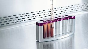
 科学海报One-Step Enrichment of Leukocyte Subsets Directly in the Blood Collection Tube
 挂图Frequencies of Immune Cells in Rat Tissue Lists the estimated frequencies of more than 15 immune cell types in Sprague Dawley rats


 沪公网安备31010102008431号