P. Petrov et al. (mar 2019)
Scientific reports 9 1 4155
Computational analysis of the evolutionarily conserved Missing In Metastasis/Metastasis Suppressor 1 gene predicts novel interactions, regulatory regions and transcriptional control.
Missing in Metastasis (MIM),or Metastasis Suppressor 1 (MTSS1),is a highly conserved protein,which links the plasma membrane to the actin cytoskeleton. MIM has been implicated in various cancers,however,its modes of action remain largely enigmatic. Here,we performed an extensive in silico characterisation of MIM to gain better understanding of its function. We detected previously unappreciated functional motifs including adaptor protein (AP) complex interaction site and a C-helix,pointing to a role in endocytosis and regulation of actin dynamics,respectively. We also identified new functional regions,characterised with phosphorylation sites or distinct hydrophilic properties. Strong negative selection during evolution,yielding high conservation of MIM,has been combined with positive selection at key sites. Interestingly,our analysis of intra-molecular co-evolution revealed potential regulatory hotspots that coincided with reduced potentially pathogenic polymorphisms. We explored databases for the mutations and expression levels of MIM in cancer. Experimentally,we focused on chronic lymphocytic leukaemia (CLL),where MIM showed high overall expression,however,downregulation on poor prognosis samples. Finally,we propose strong conservation of MTSS1 also on the transcriptional level and predict novel transcriptional regulators. Our data highlight important targets for future studies on the role of MIM in different tissues and cancers.
View Publication
产品号#:
15024
15064
产品名:
RosetteSep™人B细胞富集抗体混合物
RosetteSep™人B细胞富集抗体混合物
专家访谈
Cori Fain
Overcoming Life Circumstances to Pursue Neuroimmunology Research
Topics:
Neuroimmunology research
Immune-mediated blood brain barrier disruption in cerebral malaria
Giassi LJ et al. (AUG 2008)
Experimental biology and medicine (Maywood,N.J.) 233 8 997--1012
Expanded CD34+ human umbilical cord blood cells generate multiple lymphohematopoietic lineages in NOD-scid IL2rgamma(null) mice.
Umbilical cord blood (UCB) is increasingly being used for human hematopoietic stem cell (HSC) transplantation in children but often requires pooling multiple cords to obtain sufficient numbers for transplantation in adults. To overcome this limitation,we have used an ex vivo two-week culture system to expand the number of hematopoietic CD34(+) cells in cord blood. To assess the in vivo function of these expanded CD34(+) cells,cultured human UCB containing 1 x 10(6) CD34(+) cells were transplanted into conditioned NOD-scid IL2rgamma(null) mice. The expanded CD34(+) cells displayed short- and long-term repopulating cell activity. The cultured human cells differentiated into myeloid,B-lymphoid,and erythroid lineages,but not T lymphocytes. Administration of human recombinant TNFalpha to recipient mice immediately prior to transplantation promoted human thymocyte and T-cell development. These T cells proliferated vigorously in response to TCR cross-linking by anti-CD3 antibody. Engrafted TNFalpha-treated mice generated antibodies in response to T-dependent and T-independent immunization,which was enhanced when mice were co-treated with the B cell cytokine BLyS. Ex vivo expanded CD34(+) human UCB cells have the capacity to generate multiple hematopoietic lineages and a functional human immune system upon transplantation into TNFalpha-treated NOD-scid IL2rgamma(null) mice.
View Publication
产品号#:
09600
09650
09850
产品名:
StemSpan™ SFEM
StemSpan™ SFEM
Jones RB et al. (SEP 2009)
Journal of virology 83 17 8722--32
Human immunodeficiency virus type 1 escapes from interleukin-2-producing CD4+ T-cell responses without high-frequency fixation of mutations.
The presence of interleukin-2 (IL-2)-producing human immunodeficiency virus type 1 (HIV-1)-specific CD4(+) T-cell responses has been associated with the immunological control of HIV-1 replication; however,the causal relationship between these factors remains unclear. Here we show that IL-2-producing HIV-1-specific CD4(+) T cells can be cloned from acutely HIV-1-infected individuals. Despite the early presence of these cells,each of the individuals in the present study exhibited progressive disease,with one individual showing rapid progression. In this rapid progressor,three IL-2-producing HIV-1 Gag-specific CD4(+) T-cell responses were identified and mapped to the following optimal epitopes: HIVWASRELER,REPRGSDIAGT,and FRDYVDRFYKT. Responses to these epitopes in peripheral blood mononuclear cells were monitored longitudinally to textgreater1 year postinfection,and contemporaneous circulating plasma viruses were sequenced. A variant of the FRDYVDRFYKT epitope sequence,FRDYVDQFYKT,was observed in 1/21 plasma viruses sequenced at 5 months postinfection and 1/10 viruses at 7 months postinfection. This variant failed to stimulate the corresponding CD4(+) T-cell clone and thus constitutes an escape mutant. Responses to each of the three Gag epitopes were rapidly lost,and this loss was accompanied by a loss of antigen-specific cells in the periphery as measured by using an FRDYVDRFYKT-presenting major histocompatibility complex class II tetramer. Highly active antiretroviral therapy was associated with the reemergence of FRDYVDRFYKT-specific cells by tetramer. Thus,our data support that IL-2-producing HIV-1-specific CD4(+) T-cell responses can exert immune pressure during early HIV-1 infection but that the inability of these responses to enforce enduring control of viral replication is related to the deletion and/or dysfunction of HIV-1-specific CD4(+) T cells rather than to the fixation of escape mutations at high frequencies.
View Publication

 EasySep™小鼠TIL(CD45)正选试剂盒


 专家访谈Cori Fain Overcoming Life Circumstances to Pursue Neuroimmunology Research
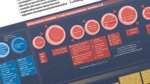 挂图T Cell Nomenclature: From Subsets to Modules A modular framework for classifying T cells by lineage, function, migration, differentiation, and antigen context.
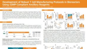 科学海报Development of Robust T Cell Manufacturing Protocols in Bioreactors Using cGMP-Compliant Ancillary Reagents


 沪公网安备31010102008431号