
 EasySep™小鼠TIL(CD45)正选试剂盒


技术资料
-

-
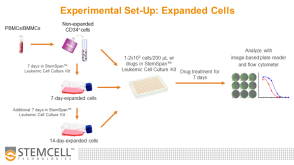
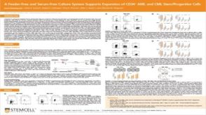 科学海报A Feeder-Free and Serum-Free Culture System Supports Expansion of CD34+ AML and CML Stem/Progenitor Cells
Conference:
ISEH 2019
发布日期: 09/10/2019 -
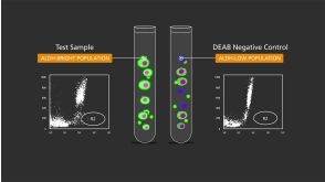
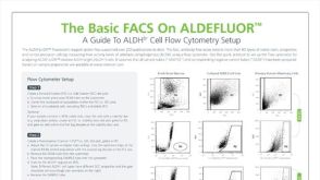 技术公告The Basic FACS on ALDEFLUOR™: The Quick Guide to Flow Cytometry
细胞类型:
乳腺细胞,前列腺细胞,癌细胞及细胞系,脑肿瘤干细胞,造血干祖细胞
发布日期: 01/23/2019 -
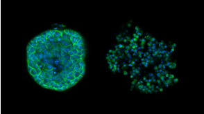
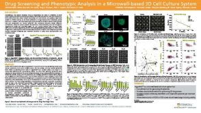 科学海报Drug Screening and Phenotypic Analysis in a Microwell-based 3D Cell Culture System
Conference:
AACR 2018
发布日期: 04/26/2018 -
-


 沪公网安备31010102008431号