Liu C et al. (MAY 2012)
Molecular biology reports 39 5 5875--81
Co-expression of Oct-4 and Nestin in human breast cancers.
The aim is to investigate the clinical implications of the Oct-4 and Nestin protein in human breast cancers. A total of 346 cases including 26 fresh and 320 paraffin-embedded tumor tissues were selected for characterizing the frequency of CD44(+)CD24(-) tumor cells by flow cytometry and the differential expression of the stem cell-related genes between CD44(+)CD24(-) and non-CD44(+)CD24(-) tumor cells was analyzed by PCR Array and immunofluorescence. In comparison with the non-CD44(+)CD24(-) tumor cells,the CD44(+)CD24(-),particularly for those with high percentage of Oct-4(+) and Nestin(+),tumor cells had higher tumorigenicity by forming mammospheres in vitro. More importantly,42 (13.125%) out of 320 tumor tissues were positive for Oct-4 and Nestin staining. Universal analysis and multivariate analysis revealed that the expression of Oct-4 and Nestin was associated significantly with younger age,pathogenic degrees,lymph node metastasis and triple-negative breast cancer independently (P textless 0.05) as well as shorter survival (P = 0.001). Oct-4 and Nestin were important regulators of the development of breast cancer,and Oct-4 and Nestin may be used as predictors for the prognosis of breast cancers.
View Publication
Taubert I et al. (APR 2011)
Cytotherapy 13 4 459--66
Characterization of hematopoietic stem cell subsets from patients with multiple myeloma after mobilization with plerixafor.
BACKGROUND AIMS: Previous studies have demonstrated that the combination of granulocyte-colony-stimulating factor (G-CSF) + plerixafor is more efficient in mobilizing CD34(+) hematopoietic stem cells (HSC) into the peripheral blood than G-CSF alone. In this study we analyzed the impact of adding plerixafor to G-CSF upon the mobilization of different HSC subsets. METHODS: We characterized the immunophenotype of HSC subsets isolated from the peripheral blood of eight patients with multiple myeloma (MM) before and after treatment with plerixafor. All patients were supposed to collect stem cells prior to high-dose chemotherapy and consecutive autologous stem cell transplantation,and therefore received front-line mobilization with 4 days of G-CSF followed by a single dose of plerixafor. Samples of peripheral blood were analyzed comparatively by flow cytometry directly before and 12 h after administration of plerixafor. RESULTS: The number of aldehyde dehydrogenase (ALDH)(bright) and CD34(+) cells was significantly higher after plerixafor treatment (1.2-5.0 and 1.5-6.0 times; both P textless 0.01) and an enrichment of the very primitive CD34(+) CD38(-) and ALDH(bright) CD34(+) CD38(-) HSC subsets was detectable. Additionally,two distinct ALDH(+) subsets could be clearly distinguished. The small ALDH(high) subset showed a higher number of CD34(+) CD38(-) cells in contrast to the total ALDH(bright) subpopulation and probably represented a very primitive subpopulation of HSC. CONCLUSIONS: A combined staining of ALDH,CD34 and CD38 might represent a powerful tool for the identification of a very rare and primitive hematopoietic stem cell subset. The addition of plerixafor mobilized not only more CD34(+) cells but was also able to increase the proportion of more primitive stem cell subsets.
View Publication
产品号#:
01700
01705
01702
产品名:
ALDEFLUOR™ 试剂盒
ALDEFLUOR™ DEAB试剂, 1.5 mM, 1 mL
ALDEFLUOR™检测缓冲液
Alison MR et al. (DEC 2010)
The Journal of pathology 222 4 335--44
Finding cancer stem cells: are aldehyde dehydrogenases fit for purpose?
Despite many years of intensive effort,there is surprisingly little consensus on the most suitable markers with which to locate and isolate stem cells from adult tissues. By comparison,the study of cancer stem cells is still in its infancy; so,unsurprisingly,there is great uncertainty as to the identity of these cells. Stem cell markers can be broadly categorized into molecular determinants of self-renewal,clonogenicity,multipotentiality,adherence to the niche,and longevity. This review assesses the utility of recognizing cancer stem cells by virtue of high expression of aldehyde dehydrogenases (ALDHs),probably significant determinants of cell survival through their ability to detoxify many potentially cytotoxic molecules,and contributing to drug resistance. Antibodies are available against the ALDH enzyme family,but the vast majority of studies have used cell sorting techniques to enrich for cells expressing these enzymes. Live cells expressing high ALDH activity are usually identified by the ALDEFLUOR kit and sorted by fluorescence activated cell sorting (FACS). For many human tumours,but notably breast cancer,cell selection based upon ALDH activity appears to be a useful marker for enriching for cells with tumour-initiating activity (presumed cancer stem cells) in immunodeficient mice,and indeed the frequency of so-called ALDH(bri) cells in many tumours can be an independent prognostic indicator.
View Publication
Kumar A et al. (JAN 2012)
Breast cancer research : BCR 14 1 R4
Evidence that GTP-binding domain but not catalytic domain of transglutaminase 2 is essential for epithelial-to-mesenchymal transition in mammary epithelial cells.
INTRODUCTION: The expression of proinflammatory protein tissue transglutaminase 2 (TG2) is frequently upregulated in multiple cancer cell types. However,the exact role of TG2 in cancer cells is not well-understood. We recently initiated studies to determine the significance of TG2 in cancer cells and observed that sustained expression of TG2 resulted in epithelial-to-mesenchymal transition (EMT) and promoted cancer stem cell (CSC) traits in mammary epithelial cells. These results suggested that TG2 could serve as a promising therapeutic target for overcoming chemoresistance and inhibiting metastatic spread of cancer cells. METHODS: Using various mutant constructs,we analyzed the activity of TG2 that is essential for promoting the EMT-CSC phenotype. RESULTS: Our results suggest that catalytically inactive TG2 (TG2-C277S) is as effective as wild-type TG2 (TG2-WT) in inducing the EMT-CSC in mammary epithelial cells. In contrast,overexpression of a GTP-binding-deficient mutant (TG2-R580A) was completely incompetent in this regard. Moreover,TG2-dependent activation of the proinflammatory transcription factor NF-κB is deemed essential for promoting the EMT-CSC phenotype in mammary epithelial cells. CONCLUSIONS: Our results suggest that the transamidation activity of TG2 is not essential for promoting its oncogenic functions and provide a strong rationale for developing small-molecule inhibitors to block GTP-binding pockets of TG2. Such inhibitors may have great potential for inhibiting the TG2-regulated pathways,reversing drug resistance and inhibiting the metastasis of cancer cells.
View Publication
产品号#:
05620
产品名:
MammoCult™ 人源培养基套装
Wu H et al. (SEP 2011)
Journal of breast cancer 14 3 175--80
Can CD44+/CD24- Tumor Cells Be Used to Determine the Extent of Breast Cancer Invasion Following Neoadjuvant Chemotherapy?
PURPOSE: To investigate the distribution of CD44(+)/CD24(-) cells in breast cancers in relation to tumor size before and after the administration of neoadjuvant chemotherapy. METHODS: CD44(+)/CD24(-) tumor cells obtained from breast cancer specimens were characterized in vivo and in vitro using tumor formation assays and mammosphere generation assays,respectively. The distribution of CD44+/CD24- tumor cells in 78 breast cancer specimens following administration of neoadjuvant chemotherapy was also evaluated using immunofluorescence assays,and this distribution was compared with the extent of tumor invasion predicted by Response Evaluation Criteria in Solid Tumours (RECIST). RESULTS: In 27/78 cases,complete remission (CR) was identified using RECIST. However,18 of these CR cases were associated with a scattered distribution of tumor stem cells in the outline of the original tumor prior to neoadjuvant chemotherapy. After neoadjuvant chemotherapy,24 cases involved cancer cells that were confined to the tumor outline,and 21 cases had tumor cells or tumor stem cells overlapping the tumor outline. In addition,there were 6 patients who were insensitive to chemotherapy,and in these cases,both cancer cells and stem cells were detected outside the contours of the tumor volume imaged prior to chemotherapy. CONCLUSION: CD44+/CD24- tumor cells may be an additional parameter to evaluate when determining the extent of breast cancer invasion.
View Publication
产品号#:
05620
产品名:
MammoCult™ 人源培养基套装
挂图
SnapShot: Breast Cancer
Overview of signaling pathways, commonly mutated genes and breast cancer subtypes

 EasySep™小鼠TIL(CD45)正选试剂盒


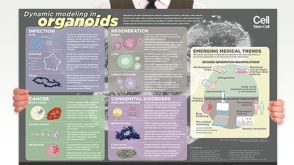 挂图Dynamic Modeling in Organoids Learn more about organoid applications for studying human health
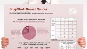 挂图SnapShot: Breast Cancer Overview of signaling pathways, commonly mutated genes and breast cancer subtypes

 沪公网安备31010102008431号