Epo receptors are not detectable in primary human tumor tissue samples.
Erythropoietin (Epo) is a cytokine that binds and activates an Epo receptor (EpoR) expressed on the surface of erythroid progenitor cells to promote erythropoiesis. While early studies suggested EpoR transcripts were expressed exclusively in the erythroid compartment,low-level EpoR transcripts were detected in nonhematopoietic tissues and tumor cell lines using sensitive RT-PCR methods. However due to the widespread use of nonspecific anti-EpoR antibodies there are conflicting data on EpoR protein expression. In tumor cell lines and normal human tissues examined with a specific and sensitive monoclonal antibody to human EpoR (A82),little/no EpoR protein was detected and it was not functional. In contrast,EpoR protein was reportedly detectable in a breast tumor cell line (MCF-7) and breast cancer tissues with an anti-EpoR polyclonal antibody (M-20),and functional responses to rHuEpo were reported with MCF-7 cells. In another study,a functional response was reported with the lung tumor cell line (NCI-H838) at physiological levels of rHuEpo. However,the specificity of M-20 is in question and the absence of appropriate negative controls raise questions about possible false-positive effects. Here we show that with A82,no EpoR protein was detectable in normal human and matching cancer tissues from breast,lung,colon,ovary and skin with little/no EpoR in MCF-7 and most other breast and lung tumor cell lines. We show further that M-20 provides false positive staining with tissues and it binds to a non-EpoR protein that migrates at the same size as EpoR with MCF-7 lysates. EpoR protein was detectable with NCI-H838 cells,but no rHuEpo-induced phosphorylation of AKT,STAT3,pS6RP or STAT5 was observed suggesting the EpoR was not functional. Taken together these results raise questions about the hypothesis that most tumors express high levels of functional EpoR protein.
View Publication
Y. Zhang et al. ( 2015)
The Journal of Immunology 194 5937-5947
Genetic Vaccines To Potentiate the Effective CD103+ Dendritic Cell-Mediated Cross-Priming of Antitumor Immunity
The development of effective cancer vaccines remains an urgent,but as yet unmet,clinical need. This deficiency is in part due to an incomplete understanding of how to best invoke dendritic cells (DC) that are crucial for the induction of tumor-specific CD8(+) T cells capable of mediating durable protective immunity. In this regard,elevated expression of the transcription factor X box-binding protein 1 (XBP1) in DC appears to play a decisive role in promoting the ability of DC to cross-present Ags to CD8(+) T cells in the therapeutic setting. Delivery of DNA vaccines encoding XBP1 and tumor Ag to skin DC resulted in increased IFN-? production by plasmacytoid DC (pDC) from skin/tumor draining lymph nodes and the cross-priming of Ag-specific CD8(+) T cell responses associated with therapeutic benefit. Antitumor protection was dependent on cross-presenting Batf3(+) DC,pDC,and CD8(+) T cells. CD103(+) DC from the skin/tumor draining lymph nodes of the immunized mice appeared responsible for activation of Ag-specific naive CD8(+) T cells,but were dependent on pDC for optimal effectiveness. Similarly,human XBP1 improved the capacity of human blood- and skin-derived DC to activate human T cells. These data support an important intrinsic role for XBP1 in DC for effective cross-priming and orchestration of Batf3(+) DC-pDC interactions,thereby enabling effective vaccine induction of protective antitumor immunity.
View Publication

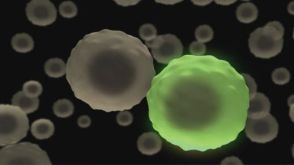
 EasySep™小鼠TIL(CD45)正选试剂盒


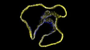


 技术公告Achieve Scalable, High-Quality Nucleic Acid Extractions with the EasySep™ Total Nucleic Acid Extraction Kit

 沪公网安备31010102008431号