Lee S-HH et al. (JUN 2000)
Nature biotechnology 18 6 675--9
Efficient generation of midbrain and hindbrain neurons from mouse embryonic stem cells.
Embryonic stem (ES) cells are clonal cell lines derived from the inner cell mass of the developing blastocyst that can proliferate extensively in vitro and are capable of adopting all the cell fates in a developing embryo. Clinical interest in the use of ES cells has been stimulated by studies showing that isolated human cells with ES properties from the inner cell mass or developing germ cells can provide a source of somatic precursors. Previous studies have defined in vitro conditions for promoting the development of specific somatic fates,specifically,hematopoietic,mesodermal,and neurectodermal. In this study,we present a method for obtaining dopaminergic (DA) and serotonergic neurons in high yield from mouse ES cells in vitro. Furthermore,we demonstrate that the ES cells can be obtained in unlimited numbers and that these neuron types are generated efficiently. We generated CNS progenitor populations from ES cells,expanded these cells and promoted their differentiation into dopaminergic and serotonergic neurons in the presence of mitogen and specific signaling molecules. The differentiation and maturation of neuronal cells was completed after mitogen withdrawal from the growth medium. This experimental system provides a powerful tool for analyzing the molecular mechanisms controlling the functions of these neurons in vitro and in vivo,and potentially for understanding and treating neurodegenerative and psychiatric diseases.
View Publication
产品号#:
06902
06952
07152
07157
00321
00322
00323
00324
00325
产品名:
N2 添加物-A
Gundemir S et al. (SEP 2016)
Neuro-Oncology now157
The complex role of transglutaminase 2 in glioblastoma proliferation
BACKGROUND Glioblastomas (GBMs) are a heterogeneous group of primary brain tumors. These tumors are resistant to therapeutic interventions and invariably recur after surgical resection. The multifunctional protein transglutaminase 2 (TG2) has been shown to promote cell survival in a number of different tumors. There is also evidence that TG2 may be a pro-survival factor in GBMs. However,the roles that TG2 plays in facilitating GBM survival and proliferation have not yet been clearly delineated . METHODS The functions of TG2 are often cell- and context-specific. Therefore,in this study we examined the ability of TG2 to facilitate GBM proliferation using colony formation assays and 5-ethynyl-2'-deoxyuridine (EdU) incorporation in several different GBM cell lines as well as neurospheres derived from patient tumors representing the 3 major subtypes of GBM tumors (mesenchymal,proneural,and classical) and maintained in the absence of serum. TG2 knockdown or selective TG2 inhibitors were used to modulate TG2 expression and activity. RESULTS We show that TG2 plays differential roles in the proliferative process depending on the cell type. In most,but not all,GBM models TG2 plays a crucial role in the proliferative process,and some but not all TG2 inhibitors were highly effective at reducing proliferation in a large subset of the GBM models. CONCLUSION Our results show that TG2 plays an important-but notoriously context-specific-role in GBM cell biology. Nonetheless,as future studies unravel the genetic fingerprints" that make TG2 inhibitors effective this information could be exploited to develop TG2 inhibitors into personalized GBM therapies.
View Publication
产品号#:
05750
05751
产品名:
NeuroCult™ NS-A 基础培养基(人)
NeuroCult™ NS-A 扩增试剂盒(人)
Zhou Y et al. (DEC 2016)
Molecular autism 7 1 42
CGG-repeat dynamics and FMR1 gene silencing in fragile X syndrome stem cells and stem cell-derived neurons.
BACKGROUND Fragile X syndrome (FXS),a common cause of intellectual disability and autism,results from the expansion of a CGG-repeat tract in the 5' untranslated region of the FMR1 gene to<200 repeats. Such expanded alleles,known as full mutation (FM) alleles,are epigenetically silenced in differentiated cells thus resulting in the loss of FMRP,a protein important for learning and memory. The timing of repeat expansion and FMR1 gene silencing is controversial. METHODS We monitored the repeat size and methylation status of FMR1 alleles with expanded CGG repeats in patient-derived induced pluripotent stem cells (iPSCs) and embryonic stem cells (ESCs) that were grown for extended period of time either as stem cells or differentiated into neurons. We used a PCR assay optimized for the amplification of large CGG repeats for sizing,and a quantitative methylation-specific PCR for the analysis of FMR1 promoter methylation. The FMR1 mRNA levels were analyzed by qRT-PCR. FMRP levels were determined by western blotting and immunofluorescence. Chromatin immunoprecipitation was used to study the association of repressive histone marks with the FMR1 gene in FXS ESCs. RESULTS We show here that while FMR1 gene silencing can be seen in FXS embryonic stem cells (ESCs),some silenced alleles contract and when the repeat number drops below ˜400,DNA methylation erodes,even when the repeat number remains<200. The resultant active alleles do not show the large step-wise expansions seen in stem cells from other repeat expansion diseases. Furthermore,there may be selection against large active alleles and these alleles do not expand further or become silenced on neuronal differentiation. CONCLUSIONS Our data support the hypotheses that (i) large expansions occur prezygotically or in the very early embryo,(ii) large unmethylated alleles may be deleterious in stem cells,(iii) methylation can occur on alleles with<400 repeats very early in embryogenesis,and (iv) expansion and contraction may occur by different mechanisms. Our data also suggest that the threshold for stable methylation of FM alleles may be higher than previously thought. A higher threshold might explain why some carriers of FM alleles escape methylation. It may also provide a simple explanation for why silencing has not been observed in mouse models with<200 repeats.
View Publication
产品号#:
05832
05850
05857
05870
05875
85850
85857
85870
85875
产品名:
STEMdiff™ 神经花环选择试剂
mTeSR™1
mTeSR™1
Marigil M et al. (JAN 2017)
PloS one 12 1 e0170501
Development of a DIPG Orthotopic Model in Mice Using an Implantable Guide-Screw System.
OBJECTIVE In this work we set to develop and to validate a new in vivo frameless orthotopic Diffuse Intrinsic Pontine Glioma (DIPG) model based in the implantation of a guide-screw system. METHODS It consisted of a guide-screw also called bolt,a Hamilton syringe with a 26-gauge needle and an insulin-like 15-gauge needle. The guide screw is 2.6 mm in length and harbors a 0.5 mm central hole which accepts the needle of the Hamilton syringe avoiding a theoretical displacement during insertion. The guide-screw is fixed on the mouse skull according to the coordinates: 1mm right to and 0.8 mm posterior to lambda. To reach the pons the Hamilton syringe is adjusted to a 6.5 mm depth using a cuff that serves as a stopper. This system allows delivering not only cells but also any kind of intratumoral chemotherapy,antibodies or gene/viral therapies. RESULTS The guide-screw was successfully implanted in 10 immunodeficient mice and the animals were inoculated with DIPG human cell lines during the same anesthetic period. All the mice developed severe neurologic symptoms and had a median overall survival of 95 days ranging the time of death from 81 to 116 days. Histopathological analysis confirmed tumor into the pons in all animals confirming the validity of this model. CONCLUSION Here we presented a reproducible and frameless DIPG model that allows for rapid evaluation of tumorigenicity and efficacy of chemotherapeutic or gene therapy products delivered intratumorally to the pons.
View Publication
产品号#:
05711
05712
05750
05751
05710
100-1281
产品名:
NeuroCult™ SM1 神经添加物
NeuroCult™ NS-A 基础培养基(人)
NeuroCult™ NS-A 扩增试剂盒(人)
NeuroCult™ SM1 神经添加物
Pollak J et al. (MAR 2017)
PLOS ONE 12 3 e0172884
Ion channel expression patterns in glioblastoma stem cells with functional and therapeutic implications for malignancy
Ion channels and transporters have increasingly recognized roles in cancer progression through the regulation of cell proliferation,migration,and death. Glioblastoma stem-like cells (GSCs) are a source of tumor formation and recurrence in glioblastoma multiforme,a highly aggressive brain cancer,suggesting that ion channel expression may be perturbed in this population. However,little is known about the expression and functional relevance of ion channels that may contribute to GSC malignancy. Using RNA sequencing,we assessed the enrichment of ion channels in GSC isolates and non-tumor neural cell types. We identified a unique set of GSC-enriched ion channels using differential expression analysis that is also associated with distinct gene mutation signatures. In support of potential clinical relevance,expression of selected GSC-enriched ion channels evaluated in human glioblastoma databases of The Cancer Genome Atlas and Ivy Glioblastoma Atlas Project correlated with patient survival times. Finally,genetic knockdown as well as pharmacological inhibition of individual or classes of GSC-enriched ion channels constrained growth of GSCs compared to normal neural stem cells. This first-in-kind global examination characterizes ion channels enriched in GSCs and explores their potential clinical relevance to glioblastoma molecular subtypes,gene mutations,survival outcomes,regional tumor expression,and experimental responses to loss-of-function. Together,the data support the potential biological and therapeutic impact of ion channels on GSC malignancy and provide strong rationale for further examination of their mechanistic and therapeutic importance.
View Publication
产品号#:
05751
70913
产品名:
NeuroCult™ NS-A 扩增试剂盒(人)
Kandasamy M et al. (MAR 2017)
Cell and Tissue Research 368 3 531--549
Glycoconjugates reveal diversity of human neural stem cells (hNSCs) derived from human induced pluripotent stem cells (hiPSCs)
Neural stem cells (NSCs) have the ability to self-renew and to differentiate into various cell types of the central nervous system. This potential can be recapitulated by human induced pluripotent stem cells (hiPSCs) in vitro. The differentiation capacity of hiPSCs is characterized by several stages with distinct morphologies and the expression of various marker molecules. We used the monoclonal antibodies (mAbs) 487(LeX),5750(LeX) and 473HD to analyze the expression pattern of particular carbohydrate motifs as potential markers at six differentiation stages of hiPSCs. Mouse ESCs were used as a comparison. At the pluripotent stage,487(LeX)-,5750(LeX)- and 473HD-related glycans were differently expressed. Later,cells of the three germ layers in embryoid bodies (hEBs) and,even after neuralization of hEBs,subpopulations of cells were labeled with these surface antibodies. At the human rosette-stage of NSCs (hR-NSC),LeX- and 473HD-related epitopes showed antibody-specific expression patterns. We also found evidence that these surface antibodies could be used to distinguish the hR-NSCs from the hSR-NSCs stages. Characterization of hNSCs(FGF-2/EGF) derived from hSR-NSCs revealed that both LeX antibodies and the 473HD antibody labeled subpopulations of hNSCs(FGF-2/EGF). Finally,we identified potential LeX carrier molecules that were spatiotemporally regulated in early and late stages of differentiation. Our study provides new insights into the regulation of glycoconjugates during early human stem cell development. The mAbs 487(LeX),5750(LeX) and 473HD are promising tools for identifying distinct stages during neural differentiation.
View Publication
产品号#:
05832
05850
05857
05870
05875
85850
85857
85870
85875
产品名:
STEMdiff™ 神经花环选择试剂
mTeSR™1
mTeSR™1
Bain G et al. (APR 1995)
Developmental biology 168 2 342--57
Embryonic stem cells express neuronal properties in vitro.
Mouse embryonic stem (ES) cells cultured as aggregates and exposed to retinoic acid are induced to express multiple phenotypes normally associated with neurons. A large percentage of treated aggregates produce a rich neuritic outgrowth. Dissociating the induced aggregates with trypsin and plating the cells as a monolayer results in cultures in which a sizable percentage of the cells have a neuronal appearance. These neuron-like cells express class III beta-tubulin and the neurofilament M subunit. Induced cultures express transcripts for neural-associated genes including the neurofilament L subunit,glutamate receptor subunits,the transcription factor Brn-3,and GFAP. Levels of neurofilament L and GAD67 and GAD65 transcripts rise dramatically upon induction. Physiological studies show that the neuron-like cells generate action potentials and express TTX-sensitive sodium channels,as well as voltage-gated potassium channels and calcium channels. We conclude that a complex system of neuronal gene expression can be activated in cultured ES cells. This system should be favorable for investigating some of the mechanisms that regulate neuronal differentiation.
View Publication
产品号#:
06902
06952
00321
00322
00323
00324
00325
产品名:
Sareen D et al. (AUG 2014)
Journal of Comparative Neurology 522 12 2707--2728
Human induced pluripotent stem cells are a novel source of neural progenitor cells (iNPCs) that migrate and integrate in the rodent spinal cord
Transplantation of human neural progenitor cells (NPCs) into the brain or spinal cord to replace lost cells,modulate the injury environment,or create a permissive milieu to protect and regenerate host neurons is a promising therapeutic strategy for neurological diseases. Deriving NPCs from human fetal tissue is feasible,although problematic issues include limited sources and ethical concerns. Here we describe a new and abundant source of NPCs derived from human induced pluripotent stem cells (iPSCs). A novel chopping technique was used to transform adherent iPSCs into free-floating spheres that were easy to maintain and were expandable (EZ spheres) (Ebert et al. [2013] Stem Cell Res 10:417–427). These EZ spheres could be differentiated towards NPC spheres with a spinal cord phenotype using a combination of all-trans retinoic acid (RA) and epidermal growth factor (EGF) and fibroblast growth factor-2 (FGF-2) mitogens. Suspension cultures of NPCs derived from human iPSCs or fetal tissue have similar characteristics,although they were not similar when grown as adherent cells. In addition,iPSC-derived NPCs (iNPCs) survived grafting into the spinal cord of athymic nude rats with no signs of overgrowth and with a very similar profile to human fetal-derived NPCs (fNPCs). These results suggest that human iNPCs behave like fNPCs and could thus be a valuable alternative for cellular regenerative therapies of neurological diseases. J. Comp. Neurol. 522:2707–2728,2014. textcopyright 2014 Wiley Periodicals,Inc.
View Publication
产品号#:
05850
05857
05870
05875
85850
85857
85870
85875
产品名:
mTeSR™1
mTeSR™1
Badizadegan K et al. (NOV 2014)
AJP: Gastrointestinal and Liver Physiology 307 10 G1002--G1012
Presence of intramucosal neuroglial cells in normal and aganglionic human colon
The enteric nervous system (ENS) is composed of neural crest-derived neurons (also known as ganglion cells) the cell bodies of which are located in the submucosal and myenteric plexuses of the intestinal wall. Intramucosal ganglion cells are known to exist but are rare and often considered ectopic. Also derived from the neural crest are enteric glial cells that populate the ganglia and the associated nerves,as well as the lamina propria of the intestinal mucosa. In Hirschsprung disease (HSCR),ganglion cells are absent from the distal gut because of a failure of neural crest-derived progenitor cells to complete their rostrocaudal migration during embryogenesis. The fate of intramucosal glial cells in human HSCR is essentially unknown. We demonstrate a network of intramucosal cells that exhibit dendritic morphology typical of neurons and glial cells. These dendritic cells are present throughout the human gut and express Tuj1,S100,glial fibrillary acidic protein,CD56,synaptophysin,and calretinin,consistent with mixed or overlapping neuroglial differentiation. The cells are present in aganglionic colon from patients with HSCR,but with an altered immunophenotype. Coexpression of Tuj1 and HNK1 in this cell population supports a neural crest origin. These findings extend and challenge the current understanding of ENS microanatomy and suggest the existence of an intramucosal population of neural crest-derived cells,present in HSCR,with overlapping immunophenotype of neurons and glia. Intramucosal neuroglial cells have not been previously recognized,and their presence in HSCR poses new questions about ENS development and the pathobiology of HSCR that merit further investigation.
View Publication
产品号#:
05750
产品名:
NeuroCult™ NS-A 基础培养基(人)
Nie S et al. (FEB 2015)
Journal of proteome research 14 2 814--22
Tenascin-C: a novel candidate marker for cancer stem cells in glioblastoma identified by tissue microarrays.
Glioblastoma multiforme (GBM) is a highly aggressive brain tumor,with dismal survival outcomes. Recently,cancer stem cells (CSCs) have been demonstrated to play a role in therapeutic resistance and are considered to be the most likely cause of cancer relapse. The identification of CSCs is an important step toward finding new and effective ways to treat GBM. Tenascin-C (TNC) protein has been identified as a potential marker for CSCs in gliomas based on previous work. Here,we have investigated the expression of TNC in tissue microarrays including 17 GBMs,18 WHO grade III astrocytomas,15 WHO grade II astrocytomas,4 WHO grade I astrocytomas,and 7 normal brain tissue samples by immunohistochemical staining. TNC expression was found to be highly associated with the grade of astrocytoma. It has a high expression level in most of the grade III astrocytomas and GBMs analyzed and a very low expression in most grade II astrocytomas,whereas it is undetectable in grade I astrocytomas and normal brain tissues. Double-immunofluorescence staining for TNC and CD133 in GBM tissues revealed that there was a high overlap between theses two positive populations. The results were further confirmed by flow cytometry analysis of TNC and CD133 in GBM-derived stem-like neurospheres in vitro. A limiting dilution assay demonstrated that the sphere formation ability of CD133(+)/TNC(+) and CD133(-)/TNC(+) cell populations is much higher than that of the CD133(+)/TNC(-) and CD133(-)/TNC(-) populations. These results suggest that TNC is not only a potential prognostic marker for GBM but also a potential marker for glioma CSCs,where the TNC(+) population is identified as a CSC population overlapping with part of the CD133(-) cell population.
View Publication

 EasySep™小鼠TIL(CD45)正选试剂盒


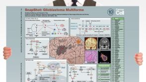 挂图SnapShot: Glioblastoma Multiforme Overview of the key concepts and mechanisms in glioblastoma multiforme biology

 沪公网安备31010102008431号