Griggs TF et al. ( 2017)
Respiratory research 18 1 84
Rhinovirus C targets ciliated airway epithelial cells.
BACKGROUND The Rhinovirus C (RV-C),first identified in 2006,produce high symptom burdens in children and asthmatics,however,their primary target host cell in the airways remains unknown. Our primary hypotheses were that RV-C target ciliated airway epithelial cells (AECs),and that cell specificity is determined by restricted and high expression of the only known RV-C cell-entry factor,cadherin related family member 3 (CDHR3). METHODS RV-C15 (C15) infection in differentiated human bronchial epithelial cell (HBEC) cultures was assessed using immunofluorescent and time-lapse epifluorescent imaging. Morphology of C15-infected differentiated AECs was assessed by immunohistochemistry. RESULTS C15 produced a scattered pattern of infection,and infected cells were shed from the epithelium. The percentage of cells infected with C15 varied from 1.4 to 14.7% depending on cell culture conditions. Infected cells had increased staining for markers of ciliated cells (acetylated-alpha-tubulin [aat],p < 0.001) but not markers of goblet cells (wheat germ agglutinin or Muc5AC,p = ns). CDHR3 expression was increased on ciliated epithelial cells,but not other epithelial cells (p < 0.01). C15 infection caused a 27.4% reduction of ciliated cells expressing CDHR3 (p < 0.01). During differentiation of AECs,CDHR3 expression progressively increased and correlated with both RV-C binding and replication. CONCLUSIONS The RV-C only replicate in ciliated AECs in vitro,leading to infected cell shedding. CDHR3 expression positively correlates with RV-C binding and replication,and is largely confined to ciliated AECs. Our data imply that factors regulating differentiation and CDHR3 production may be important determinants of RV-C illness severity.
View Publication
产品号#:
05001
05021
05022
产品名:
PneumaCult™-ALI 培养基
PneumaCult™-ALI 培养基含12 mm Transwell®插件
PneumaCult™-ALI 培养基含6.5 mm Transwell®插件
Herawati E et al. ( 2016)
Journal of Cell Biology 214 5 571--586
Multiciliated cell basal bodies align in stereotypical patterns coordinated by the apical cytoskeleton
Multiciliated cells (MCCs) promote fluid flow through coordinated ciliary beating,which requires properly organized basal bodies (BBs). Airway MCCs have large numbers of BBs,which are uniformly oriented and,as we show here,align linearly. The mechanism for BB alignment is unexplored. To study this mechanism,we developed a long-term and high-resolution live-imaging system and used it to observe green fluorescent protein"centrin2"labeled BBs in cultured mouse tracheal MCCs. During MCC differentiation,the BB array adopted four stereotypical patterns,from a clustering floret? pattern to the linear alignment.? This alignment process was correlated with BB orientations,revealed by double immunostaining for BBs and their asymmetrically associated basal feet (BF). The BB alignment was disrupted by disturbing apical microtubules with nocodazole and by a BF-depleting Odf2 mutation. We constructed a theoretical model,which indicated that the apical cytoskeleton,acting like a viscoelastic fluid,provides a self-organizing mechanism in tracheal MCCs to align BBs linearly for mucociliary transport.
View Publication
产品号#:
05001
05021
05022
05008
产品名:
PneumaCult™-ALI 培养基
PneumaCult™-ALI 培养基含12 mm Transwell®插件
PneumaCult™-ALI 培养基含6.5 mm Transwell®插件
PneumaCult™-Ex 培养基
Cao X et al. (JAN 2015)
Respiratory research 16 30
Tight junction disruption by cadmium in an in vitro human airway tissue model.
BACKGROUND: The cadmium (Cd) present in air pollutants and cigarette smoke has the potential of causing multiple adverse health outcomes involving damage to pulmonary and cardiovascular tissue. Injury to pulmonary epithelium may include alterations in tight junction (TJ) integrity,resulting in impaired epithelial barrier function and enhanced penetration of chemicals and biomolecules. Herein,we investigated mechanisms involved in the disruption of TJ integrity by Cd exposure using an in vitro human air-liquid-interface (ALI) airway tissue model derived from normal primary human bronchial epithelial cells. METHODS: ALI cultures were exposed to noncytotoxic doses of CdCl2 basolaterally and TJ integrity was measured by Trans-Epithelial Electrical Resistance (TEER) and immunofluorescence staining with TJ markers. PCR array analysis was used to identify genes involved with TJ collapse. To explore the involvement of kinase signaling pathways,cultures were treated with CdCl2 in the presence of kinase inhibitors specific for cellular Src or Protein Kinase C (PKC). RESULTS: Noncytotoxic doses of CdCl2 resulted in the collapse of barrier function,as demonstrated by TEER measurements and Zonula occludens-1 (ZO-1) and occludin staining. CdCl2 exposure altered the expression of several groups of genes encoding proteins involved in TJ homeostasis. In particular,down-regulation of select junction-interacting proteins suggested that a possible mechanism for Cd toxicity involves disruption of the peripheral junctional complexes implicated in connecting membrane-bound TJ components to the actin cytoskeleton. Inhibition of kinase signaling using inhibitors specific for cellular Src or PKC preserved the integrity of TJs,possibly by preventing occludin tyrosine hyperphosphorylation,rather than reversing the down-regulation of the junction-interacting proteins. CONCLUSIONS: Our findings indicate that acute doses of Cd likely disrupt TJ integrity in human ALI airway cultures both through occludin hyperphosphorylation via kinase activation and by direct disruption of the junction-interacting complex.
View Publication
Ishikawa S et al. ( 2017)
Respiratory Research 18 1 1--11
A 3D epithelial-mesenchymal co-culture model of human bronchial tissue recapitulates multiple features of airway tissue remodeling by TGF-β1 treatment
BACKGROUND: The collagen gel contraction assay measures gel size to assess the contraction of cells embedded in collagen gel matrices. Using the assay with lung fibroblasts is useful in studying the lung tissue remodeling process in wound healing and disease development. However,the involvement of bronchial epithelial cells in this process should also be investigated. METHODS: We applied a layer of mucociliary differentiated bronchial epithelial cells onto collagen gel matrices with lung fibroblasts. This co-culture model enables direct contact between epithelial and mesenchymal cells. We stimulated the culture with transforming growth factor (TGF) beta1 as an inducer of tissue remodeling for 21 days,and measured gel size,histological changes,and expression of factors related to extracellular matrix homeostasis. RESULTS: TGF-beta1 exerted a concentration-dependent effect on collagen gel contraction and on contractile myofibroblasts in the mesenchymal collagen layer. TGF-beta1 also induced expression of the mesenchymal marker vimentin in the basal layer of the epithelium,suggesting the induction of epithelial-mesenchymal transition. In addition,the expression of various genes encoding extracellular matrix proteins was upregulated. Fibrotic tenascin-C accumulated in the sub-epithelial region of the co-culture model. CONCLUSION: Our findings indicate that TGF-beta1 can affect both epithelial and mesenchymal cells,and induce gel contraction and structural changes. Our novel in vitro co-culture model will be a useful tool for investigating the roles of epithelial cells,fibroblasts,and their interactions in the airway remodeling process.
View Publication
Efficient Derivation of Functional Human Airway Epithelium from Pluripotent Stem Cells via Temporal Regulation of Wnt Signaling.
Effective derivation of functional airway organoids from induced pluripotent stem cells (iPSCs) would provide valuable models of lung disease and facilitate precision therapies for airway disorders such as cystic fibrosis. However,limited understanding of human airway patterning has made this goal challenging. Here,we show that cyclical modulation of the canonical Wnt signaling pathway enables rapid directed differentiation of human iPSCs via an NKX2-1+progenitor intermediate into functional proximal airway organoids. We find that human NKX2-1+progenitors have high levels of Wnt activation but respond intrinsically to decreases in Wnt signaling by rapidly patterning into proximal airway lineages at the expense of distal fates. Using this directed approach,we were able to generate cystic fibrosis patient-specific iPSC-derived airway organoids with a defect in forskolin-induced swelling that is rescued by gene editing to correct the disease mutation. Our approach has many potential applications in modeling and drug screening for airway diseases.
View Publication
产品号#:
05001
05021
05022
产品名:
PneumaCult™-ALI 培养基
PneumaCult™-ALI 培养基含12 mm Transwell®插件
PneumaCult™-ALI 培养基含6.5 mm Transwell®插件
Shikotra A et al. ( 2017)
Journal of immunology (Baltimore,Md. : 1950) 198 8 3307--3317
A CEACAM6-High Airway Neutrophil Phenotype and CEACAM6-High Epithelial Cells Are Features of Severe Asthma.
Severe asthma represents a major unmet clinical need; understanding the pathophysiology is essential for the development of new therapies. Using microarray analysis,we previously found three immunological clusters in asthma: Th2-high,Th17-high,and Th2/17-low. Although new therapies are emerging for Th2-high disease,identifying molecular pathways in Th2-low disease remains an important goal. Further interrogation of our previously described microarray dataset revealed upregulation of gene expression for carcinoembryonic Ag cell adhesion molecule (CEACAM) family members in the bronchi of patients with severe asthma. Our aim was therefore to explore the distribution and cellular localization of CEACAM6 using immunohistochemistry on bronchial biopsy tissue obtained from patients with mild-to-severe asthma and healthy control subjects. Human bronchial epithelial cells were used to investigate cytokine and corticosteroid in vitro regulation of CEACAM6 gene expression. CEACAM6 protein expression in bronchial biopsies was increased in airway epithelial cells and lamina propria inflammatory cells in severe asthma compared with healthy control subjects. CEACAM6 in the lamina propria was localized to neutrophils predominantly. Neutrophil density in the bronchial mucosa was similar across health and the spectrum of asthma severity,but the percentage of neutrophils expressing CEACAM6 was significantly increased in severe asthma,suggesting the presence of an altered neutrophil phenotype. CEACAM6 gene expression in cultured epithelial cells was upregulated by wounding and neutrophil elastase. In summary,CEACAM6 expression is increased in severe asthma and primarily associated with airway epithelial cells and tissue neutrophils. CEACAM6 may contribute to the pathology of treatment-resistant asthma via neutrophil and airway epithelial cell-dependent pathways.
View Publication
产品号#:
05001
05021
05022
产品名:
PneumaCult™-ALI 培养基
PneumaCult™-ALI 培养基含12 mm Transwell®插件
PneumaCult™-ALI 培养基含6.5 mm Transwell®插件
Barkal LJ et al. ( 2017)
Nature Communications 8 1
Microbial volatile communication in human organotypic lung models
We inhale respiratory pathogens continuously,and the subsequent signaling events between host and microbe are complex,ultimately resulting in clearance of the microbe,stable colonization of the host,or active disease. Traditional in vitro methods are ill-equipped to study these critical events in the context of the lung microenvironment. Here we introduce a microscale organotypic model of the human bronchiole for studying pulmonary infection. By leveraging microscale techniques,the model is designed to approximate the structure of the human bronchiole,containing airway,vascular,and extracellular matrix compartments. To complement direct infection of the organotypic bronchiole,we present a clickable extension that facilitates volatile compound communication between microbial populations and the host model. Using Aspergillus fumigatus,a respiratory pathogen,we characterize the inflammatory response of the organotypic bronchiole to infection. Finally,we demonstrate multikingdom,volatile-mediated communication between the organotypic bronchiole and cultures of Aspergillus fumigatus and Pseudomonas aeruginosa.
View Publication
产品号#:
05001
05021
05022
产品名:
PneumaCult™-ALI 培养基
PneumaCult™-ALI 培养基含12 mm Transwell®插件
PneumaCult™-ALI 培养基含6.5 mm Transwell®插件
Nikoli&cacute et al. ( 2017)
eLife 6 1--33
Human embryonic lung epithelial tips are multipotent progenitors that can be expanded in vitro as long-term self-renewing organoids
The embryonic mouse lung is a widely used substitute for human lung development. For example,attempts to differentiate human pluripotent stem cells to lung epithelium rely on passing through progenitor states that have only been described in mouse. The tip epithelium of the branching mouse lung is a multipotent progenitor pool that self-renews and produces differentiating descendants. We hypothesized that the human distal tip epithelium is an analogous progenitor population and tested this by examining morphology,gene expression and in vitro self-renewal and differentiation capacity of human tips. These experiments confirm that human and mouse tips are analogous and identify signalling pathways that are sufficient for long-term self-renewal of human tips as differentiation-competent organoids. Moreover,we identify mouse-human differences,including markers that define progenitor states and signalling requirements for long-term self-renewal. Our organoid system provides a genetically-tractable tool that will allow these human-specific features of lung development to be investigated.
View Publication
产品号#:
05001
05021
05022
产品名:
PneumaCult™-ALI 培养基
PneumaCult™-ALI 培养基含12 mm Transwell®插件
PneumaCult™-ALI 培养基含6.5 mm Transwell®插件
Ahmadi S et al. ( 2017)
npj Genomic Medicine 2 1 12
Phenotypic profiling of CFTR modulators in patient-derived respiratory epithelia
Pulmonary disease is the major cause of morbidity and mortality in patients with cystic fibrosis,a disease caused by mutations in the Cystic Fibrosis Transmembrane conductance Regulator (CFTR) gene. Heterogeneity in CFTR genotype-phenotype relationships in affected individuals plus the escalation of drug discovery targeting specific mutations highlights the need to develop robust in vitro platforms with which to stratify therapeutic options using relevant tissue. Toward this goal,we adapted a fluorescence plate reader assay of apical CFTR-mediated chloride conductance to enable profiling of a panel of modulators on primary nasal epithelial cultures derived from patients bearing different CFTR mutations. This platform faithfully recapitulated patient-specific responses previously observed in the gold-standard but relatively low-throughput Ussing chamber. Moreover using this approach we identified a novel strategy with which to augment the response to an approved drug in specific patients. In proof of concept studies we also validated the use of this platform in measuring drug responses in lung cultures differentiated from cystic fibrosis iPS cells. Taken together we show that this medium throughput assay of CFTR activity has the potential to stratify cystic fibrosis patient-specific responses to approved drugs and investigational compounds in vitro in primary and iPS cell-derived airway cultures.
View Publication
产品号#:
05001
05021
05022
产品名:
PneumaCult™-ALI 培养基
PneumaCult™-ALI 培养基含12 mm Transwell®插件
PneumaCult™-ALI 培养基含6.5 mm Transwell®插件
Li X et al. (AUG 2012)
Journal of thoracic oncology : official publication of the International Association for the Study of Lung Cancer 7 8 1235--45
Aldehyde dehydrogenase 1A1 possesses stem-like properties and predicts lung cancer patient outcome.
INTRODUCTION: Lung cancer contains a small population of cancer stem cells that contribute to its initiation and progression. We investigated the biological function and clinical significance of aldehyde dehydrogenase 1A1 (ALDH1A1) in non-small-cell lung carcinoma (NSCLC). METHODS: ALDH1A1 assay or small interfering RNA transfection was employed to isolate ALDH1A1+ cells or knock down ALDH1A1 expression in H2087 cells,respectively. Biological functions of ALDH1A1+ and ALDH1A1 silenced cells were investigated using in vitro and in vivo methods. ALDH1A1 expression was analyzed using immunohistochemistry on tissue microarrays with 179 lung cancer tissues and 26 normal lung tissues. RESULTS: The abilities of clone formation,proliferation,cell growth,and migration were increased in ALDH1A1+ and ALDH1A1 silenced cells. ALDH1A1+ lung cancer cells initiated tumors that resembled the histopathologic characteristics and heterogeneity of the parental lung cancer cells in mice. The silencing of ALDH1A1 expression in H2087 lung cancer cells inhibited cell proliferation and migration significantly. ALDH1A1 was expressed in 42% of normal lung tissues (11 of 26),with strong expression in the basal cells and globular cells of the normal bronchus and weak expression in the alveolar epithelial cells. Compared with normal lung tissues,45% of NSCLC samples (81 of 179) were read as positive for ALDH1A1. Positive ALDH1A1 expression was correlated with patients' smoking status (p = 0.022),lymph-node metastasis (p = 0.006),clinical stage (p = 0.004),and a decreased overall survival time (p textless 0.001). Positive ALDH1A1 expression in lung cancer tissues was an independent prognostic factor for NSCLC (odds ratio = 5.232,p textless 0.001). CONCLUSION: Elucidating the biological functions of ALDH1A1 could be helpful in studying lung tumorigenesis and for developing new therapeutic approaches.
View Publication
产品号#:
01700
01705
01702
产品名:
ALDEFLUOR™ 试剂盒
ALDEFLUOR™ DEAB试剂, 1.5 mM, 1 mL
ALDEFLUOR™检测缓冲液
挂图
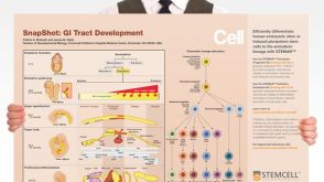
SnapShot: GI Tract Development
Overview of gastrointestinal tract specification signals and summary of pancreatic cellular hierarchy and cell markers

 EasySep™小鼠TIL(CD45)正选试剂盒


 挂图SnapShot: GI Tract Development Overview of gastrointestinal tract specification signals and summary of pancreatic cellular hierarchy and cell markers
 沪公网安备31010102008431号