Ma I and Allan AL (JUN 2011)
Stem cell reviews 7 2 292--306
The role of human aldehyde dehydrogenase in normal and cancer stem cells.
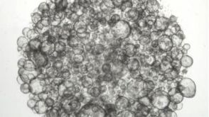
Normal stem cells and cancer stem cells (CSCs) share similar properties,in that both have the capacity to self-renew and differentiate into multiple cell types. In both the normal stem cell and cancer stem cell fields,there has been a great need for a universal marker that can effectively identify and isolate these rare populations of cells in order to characterize them and use this information for research and therapeutic purposes. Currently,it would appear that certain isoenzymes of the aldehyde dehydrogenase (ALDH) superfamily may be able to fulfill this role as a marker for both normal and cancer stem cells. ALDH has been identified as an important enzyme in the protection of normal hematopoietic stem cells,and is now also widely used as a marker to identify and isolate various types of normal stem cells and CSCs. In addition,emerging evidence suggests that ALDH1 is not only a marker for stem cells,but may also play important functional roles related to self-protection,differentiation,and expansion. This comprehensive review discusses the role that ALDH plays in normal stem cells and CSCs,with focus on ALDH1 and ALDH3A1. Discrepancies in the functional themes between cell types and future perspectives for therapeutic applications will also be discussed.
View Publication
产品号#:
01700
01705
01702
产品名:
ALDEFLUOR™ 试剂盒
ALDEFLUOR™ DEAB试剂, 1.5 mM, 1 mL
ALDEFLUOR™检测缓冲液
Karki R et al. (DEC 2016)
Nature
NLRC3 is an inhibitory sensor of PI3K-mTOR pathways in cancer.
NLRs (nucleotide-binding domain and leucine-rich repeats) belong to a large family of cytoplasmic sensors that regulate an extraordinarily diverse range of biological functions. One of these functions is to contribute to immunity against infectious diseases,but dysregulation of their functional activity leads to the development of inflammatory and autoimmune diseases. Cytoplasmic innate immune sensors,including NLRs,are central regulators of intestinal homeostasis. NLRC3 (also known as CLR16.2 or NOD3) is a poorly characterized member of the NLR family and was identified in a genomic screen for genes encoding proteins bearing leucine-rich repeats (LRRs) and nucleotide-binding domains. Expression of NLRC3 is drastically reduced in the tumour tissue of patients with colorectal cancer compared to healthy tissues,highlighting an undefined potential function for this sensor in the development of cancer. Here we show that mice lacking NLRC3 are hyper-susceptible to colitis and colorectal tumorigenesis. The effect of NLRC3 is most dominant in enterocytes,in which it suppresses activation of the mTOR signalling pathways and inhibits cellular proliferation and stem-cell-derived organoid formation. NLRC3 associates with PI3Ks and blocks activation of the PI3K-dependent kinase AKT following binding of growth factor receptors or Toll-like receptor 4. These findings reveal a key role for NLRC3 as an inhibitor of the mTOR pathways,mediating protection against colorectal cancer.
View Publication
产品号#:
06005
产品名:
IntestiCult™ 类器官生长培养基 (小鼠)
Okkelman IA et al. ( 2016)
PloS one 11 12 e0167385
Use of Fluorescence Lifetime Imaging Microscopy (FLIM) as a Timer of Cell Cycle S Phase.
Incorporation of thymidine analogues in replicating DNA,coupled with antibody and fluorophore staining,allows analysis of cell proliferation,but is currently limited to monolayer cultures,fixed cells and end-point assays. We describe a simple microscopy imaging method for live real-time analysis of cell proliferation,S phase progression over several division cycles,effects of anti-proliferative drugs and other applications. It is based on the prominent (˜ 1.7-fold) quenching of fluorescence lifetime of a common cell-permeable nuclear stain,Hoechst 33342 upon the incorporation of 5-bromo-2'-deoxyuridine (BrdU) in genomic DNA and detection by fluorescence lifetime imaging microscopy (FLIM). We show that quantitative and accurate FLIM technique allows high-content,multi-parametric dynamic analyses,far superior to the intensity-based imaging. We demonstrate its uses with monolayer cell cultures,complex 3D tissue models of tumor cell spheroids and intestinal organoids,and in physiological study with metformin treatment.
View Publication
产品号#:
06005
产品名:
IntestiCult™ 类器官生长培养基 (小鼠)
Rong S et al. (JUN 2017)
Journal of lipid research jlr.M077610
Cholesterol auxotrophy and intolerance to ezetimibe in mice with SREBP-2 deficiency in the intestine.
Sterol regulatory element-binding protein-2 (SREBP-2) activates transcription of all genes needed for cholesterol biosynthesis. To study SREBP-2 function in intestine,we generated a mouse model (Vil-BP2(-/-) ) in which Cre recombinase ablates SREBP-2 in intestinal epithelia. Intestines of Vil-BP2(-/-) mice had reduced expression of genes required for sterol synthesis,in vivo sterol synthesis rates,and epithelial cholesterol contents. On a cholesterol-free diet,they displayed chronic enteropathy with histological abnormalities of both villi and crypts,growth restriction,and reduced survival that was prevented by supplementation of cholesterol in the diet. Likewise,SREBP-2-deficient enteroids required exogenous cholesterol for growth. Blockade of luminal cholesterol uptake into enterocytes with ezetimibe precipitated acutely lethal intestinal damage in Vil-BP2(-/-) mice,highlighting the critical interplay in the small intestine of sterol absorption via NPC1L1 and sterol synthesis via SREBP-2 in sustaining the intestinal mucosa. These data show that small intestine requires SREBP-2 to drive cholesterol synthesis that sustains the intestinal epithelia when uptake of cholesterol from the gut lumen is not available,and provide a unique example of cholesterol auxotrophy expressed in an intact,adult mammal.
View Publication
产品号#:
06005
产品名:
IntestiCult™ 类器官生长培养基 (小鼠)
Aladegbami B et al. (JUL 2017)
Scientific reports 7 1 5580
Epithelial cell specific Raptor is required for initiation of type 2 mucosal immunity in small intestine.
Intestinal tuft cells are one of 4 secretory cell linages in the small intestine and the source of IL-25,a critical initiator of the type 2 immune response to parasite infection. When Raptor,a critical scaffold protein for mammalian target of rapamycin complex 1 (mTORC1),was acutely deleted in intestinal epithelium via Tamoxifen injection in Tritrichomonas muris (Tm) infected mice,tuft cells,IL-25 in epithelium and IL-13 in the mesenchyme were significantly reduced,but Tm burden was not affected. When Tm infected mice were treated with rapamycin,DCLK1 and IL-25 expression in enterocytes and IL-13 expression in mesenchyme were diminished. After massive small bowel resection,tuft cells and Tm were diminished due to the diet used postoperatively. The elimination of Tm and subsequent re-infection of mice with Tm led to type 2 immune response only in WT,but Tm colonization in both WT and Raptor deficient mice. When intestinal organoids were stimulated with IL-4,tuft cells and IL-25 were induced in both WT and Raptor deficient organoids. In summary,our study reveals that enterocyte specific Raptor is required for initiating a type 2 immune response which appears to function through the regulation of mTORC1 activity.
View Publication
A. Stern et al. (Apr 2022)
SLAS Discovery 27 201-208
The CellRaft AIR? system: A novel system enabling organoid imaging, identification, and isolation
Three-dimensional (3D) culture systems have been developed that can re-capitulate organ level responses,simulate compound diffusion through complex structures,and assess cellular heterogeneity of tissues,making them attractive models for advanced in vitro research and discovery. Organoids are a unique subtype of 3D cell culture that are grown from stem cells,are self-organizing,and closely replicate in vivo pathophysiology. Organoids have been used to understand tissue development,model diseases,test drug sensitivity and toxicity,and advance regenerative medicine. However,traditional organoid culture methods are inadequate because they are low throughput and ill-suited for single organoid imaging,phenotypic assessment,and isolation from heterogenous organoid populations. To address these bottlenecks,we have adapted our tissue culture consumable and instrumentation to enable automated imaging,identification,and isolation of individual organoids. Organoids grown on the 3D CytoSort? Array can be reliably tracked,imaged,and phenotypically analyzed using brightfield and fluorescent microscopy as they grow over time,then released and transferred fully intact for use in downstream applications. Using mouse hepatic and pancreatic organoids,we have demonstrated the use of this technology for single-organoid imaging,clonal organoid generation,parent organoid subcloning,and single-organoid RNA extraction for downstream gene expression or transcriptomic analysis. The results validate the ability of the CellRaft AIR? System to facilitate efficient,user-friendly,and automated workflows broadly applicable to organoid research by overcoming several pain points: 1) single organoid time-course imaging and phenotypic assessment,2) establishment of single cell-derived organoids,and 3) isolation and retrieval of single organoids for downstream applications.
View Publication

 EasySep™小鼠TIL(CD45)正选试剂盒


 沪公网安备31010102008431号