Sulforaphane targets pancreatic tumour-initiating cells by NF-kappaB-induced antiapoptotic signalling.
BACKGROUND AND AIMS: Emerging evidence suggests that highly treatment-resistant tumour-initiating cells (TICs) play a central role in the pathogenesis of pancreatic cancer. Tumour necrosis factor-related apoptosis-inducing ligand (TRAIL) is considered to be a novel anticancer agent; however,recent studies have shown that many pancreatic cancer cells are resistant to apoptosis induction by TRAIL due to TRAIL-activated nuclear factor-kappaB (NF-kappaB) signalling. Several chemopreventive agents are able to inhibit NF-kappaB,and favourable results have been obtained--for example,for the broccoli compound sulforaphane-in preventing metastasis in clinical studies. The aim of the study was to identify TICs in pancreatic carcinoma for analysis of resistance mechanisms and for definition of sensitising agents. METHODS: TICs were defined by expression patterns of a CD44(+)/CD24(-),CD44(+)/CD24(+) or CD44(+)/CD133(+) phenotype and correlation to growth in immunodeficient mice,differentiation grade,clonogenic growth,sphere formation,aldehyde dehydrogenase (ALDH) activity and therapy resistance. RESULTS: Mechanistically,specific binding of transcriptionally active cRel-containing NF-kappaB complexes in TICs was observed. Sulforaphane prevented NF-kappaB binding,downregulated apoptosis inhibitors and induced apoptosis,together with prevention of clonogenicity. Gemcitabine,the chemopreventive agents resveratrol and wogonin,and the death ligand TRAIL were less effective. In a xenograft model,sulforaphane strongly blocked tumour growth and angiogenesis,while combination with TRAIL had an additive effect without obvious cytotoxicity in normal cells. Freshly isolated patient tumour cells expressing markers for TICs could be sensitised by sulforaphane for TRAIL-induced cytotoxicity. CONCLUSION: The data provide new insights into resistance mechanisms of TICs and suggest the combination of sulforaphane with TRAIL as a promising strategy for targeting of pancreatic TICs.
View Publication
Ben-David U et al. (SEP 2014)
Nature communications 5 4825
Aneuploidy induces profound changes in gene expression, proliferation and tumorigenicity of human pluripotent stem cells.
Human pluripotent stem cells (hPSCs) tend to acquire genomic aberrations in culture,the most common of which is trisomy of chromosome 12. Here we dissect the cellular and molecular implications of this trisomy in hPSCs. Global gene expression analyses reveal that trisomy 12 profoundly affects the gene expression profile of hPSCs,inducing a transcriptional programme similar to that of germ cell tumours. Comparison of proliferation,differentiation and apoptosis between diploid and aneuploid hPSCs shows that trisomy 12 significantly increases the proliferation rate of hPSCs,mainly as a consequence of increased replication. Furthermore,trisomy 12 increases the tumorigenicity of hPSCs in vivo,inducing transcriptionally distinct teratomas from which pluripotent cells can be recovered. Last,a chemical screen of 89 anticancer drugs discovers that trisomy 12 raises the sensitivity of hPSCs to several replication inhibitors. Together,these findings demonstrate the extensive effect of trisomy 12 and highlight its perils for successful hPSC applications.
View Publication
产品号#:
05850
05857
05870
05875
07909
85850
85857
85870
85875
产品名:
IV型胶原酶(1mg /mL)
mTeSR™1
mTeSR™1
Vanden Bempt M et al. (MAR 2016)
Leukemia March 8 Epub ahead of print
Generation of the Fip1l1–Pdgfra fusion gene using CRISPR/Cas genome editing
Calcagno AM et al. (NOV 2010)
Journal of the National Cancer Institute 102 21 1637--52
Prolonged drug selection of breast cancer cells and enrichment of cancer stem cell characteristics.
BACKGROUND: Cancer stem cells are presumed to have virtually unlimited proliferative and self-renewal abilities and to be highly resistant to chemotherapy,a feature that is associated with overexpression of ATP-binding cassette transporters. We investigated whether prolonged continuous selection of cells for drug resistance enriches cultures for cancer stem-like cells. METHODS: Cancer stem cells were defined as CD44+/CD24�?� cells that could self-renew (ie,generate cells with the tumorigenic CD44+/CD24�?� phenotype),differentiate,invade,and form tumors in vivo. We used doxorubicin-selected MCF-7/ADR cells,weakly tumorigenic parental MCF-7 cells,and MCF-7/MDR,an MCF-7 subline with forced expression of ABCB1 protein. Cells were examined for cell surface markers and side-population fractions by microarray and flow cytometry,with in vitro invasion assays,and for ability to form mammospheres. Xenograft tumors were generated in mice to examine tumorigenicity (n = 52). The mRNA expression of multidrug resistance genes was examined in putative cancer stem cells and pathway analysis of statistically significantly differentially expressed genes was performed. All statistical tests were two-sided. RESULTS: Pathway analysis showed that MCF-7/ADR cells express mRNAs from ABCB1 and other genes also found in breast cancer stem cells (eg,CD44,TGFB1,and SNAI1). MCF-7/ADR cells were highly invasive,formed mammospheres,and were tumorigenic in mice. In contrast to parental MCF-7 cells,more than 30% of MCF-7/ADR cells had a CD44+/CD24�?� phenotype,could self-renew,and differentiate (ie,produce CD44+/CD24�?� and CD44+/CD24+ cells) and overexpressed various multidrug resistance-linked genes (including ABCB1,CCNE1,and MMP9). MCF-7/ADR cells were statistically significantly more invasive in Matrigel than parental MCF-7 cells (MCF-7 cells = 0.82 cell per field and MCF-7/ADR = 7.51 cells per field,difference = 6.69 cells per field,95% confidence interval = 4.82 to 8.55 cells per field,P textless .001). No enrichment in the CD44+/CD24�?� or CD133+ population was detected in MCF-7/MDR. CONCLUSION: The cell population with cancer stem cell characteristics increased after prolonged continuous selection for doxorubicin resistance.
View Publication
产品号#:
01700
01705
01702
产品名:
ALDEFLUOR™ 试剂盒
ALDEFLUOR™ DEAB试剂, 1.5 mM, 1 mL
ALDEFLUOR™检测缓冲液
Weidanz Ja et al. (OCT 2006)
Journal of Immunology (Baltimore,Md. : 1950) 177 8 5088--97
Levels of specific peptide-HLA class I complex predicts tumor cell susceptibility to CTL killing.
Recognition of tumor-associated Ags (TAAs) on tumor cells by CTLs and the subsequent tumor cell death are assumed to be dependent on TAA protein expression and to correlate directly with the level of peptide displayed in the binding site of the HLA class I molecule. In this study we evaluated whether the levels of Her-2/neu protein expression on human tumor cell lines directly correlate with HLA-A*0201/Her2/neu peptide presentation and CTL recognition. We developed a TCR mimic (TCRm) mAb designated 1B8 that specifically recognizes the HLA-A2.1/Her2/neu peptide (369-377) (Her2(369)-A2) complex. TCRm mAb staining intensity varied for the five human tumor cell lines analyzed,suggesting quantitative differences in levels of the Her2(369)-A2 complex on these cells. Analysis of tumor cell lines pretreated with IFN-gamma and TNF-alpha for Her2/neu protein and HLA-A2 molecule expression did not reveal a direct correlation between the levels of Her2/neu Ag,HLA-A2 molecule,and Her2(369)-A2 complex expression. However,compared with untreated cells,cytokine-treated cell lines showed an increase in Her2(369)-A2 epitope density that directly correlated with enhanced tumor cell death (p = 0.05). Although a trend was observed between tumor cell lysis and the level of the Her2(369)-A2 complex for untreated cells,the association was not significant. These findings suggest that tumor cell susceptibility to CTL-mediated lysis may be predicted based on the level of specific peptide-MHC class I expression rather than on the total level of TAA expression. Further,these studies demonstrate the potential of the TCRm mAb for validation of endogenous HLA-peptide epitopes on tumor cells.
View Publication
产品号#:
03800
03801
03802
03803
03804
03805
03806
03831
产品名:
ClonaCell™-HY杂交瘤试剂盒
ClonaCell™-HY培养基A
ClonaCell™-HY 培养基 B
ClonaCell™-HY 培养基 C
ClonaCell™-HY 培养基 D
ClonaCell™-HY 培养基 E
ClonaCell™-HY PEG
ClonaCell™-HY 液体 HAT 筛选培养基
Kern J et al. (OCT 2009)
Blood 114 18 3960--7
GRP-78 secreted by tumor cells blocks the antiangiogenic activity of bortezomib.
Antiangiogenic effects of the proteasome inhibitor bortezomib were analyzed on tumor xenografts in vivo. Bortezomib strongly inhibited angiogenesis and vascularization in the chicken chorioallantoic membrane. Bortezomib's inhibitory effects on chorioallantoic membrane vascularization were abrogated in the presence of distinct tumor xenografts,thanks to a soluble factor secreted by tumor cells. Through size-exclusion and ion-exchange chromatography as well as mass spectroscopy,we identified GRP-78,a chaperone protein of the unfolded protein response,as being responsible for bortezomib resistance. Indeed,a variety of bortezomib-resistant solid tumor cell lines (PC-3,HRT-18),but not myeloma cell lines (U266,OPM-2),were able to secrete high amounts of GRP-78. Recombinant GRP-78 conferred bortezomib resistance to endothelial cells and OPM-2 myeloma cells. Knockdown of GRP78 gene expression in tumor cells and immunodepletion of GRP-78 protein from tumor cell supernatants restored bortezomib sensitivity. GRP-78 did not bind or complex bortezomib but induced prosurvival signals by phosphorylation of extracellular signal-related kinase and inhibited p53-mediated expression of proapoptotic Bok and Noxa proteins in endothelial cells. From our data,we conclude that distinct solid tumor cells are able to secrete GRP-78 into the tumor microenvironment,thus demonstrating a hitherto unknown mechanism of resistance to bortezomib.
View Publication
Wittman VP et al. (SEP 2006)
The Journal of Immunology 177 6 4187--95
Antibody targeting to acClass I MHC-peptide epitope promotes tumor cell death
Therapeutic mAbs that target tumor-associated Ags on the surface of malignant cells have proven to be an effective and specific option for the treatment of certain cancers. However,many of these protein markers of carcinogenesis are not expressed on the cells' surface. Instead these tumor-associated Ags are processed into peptides that are presented at the cell surface,in the context of MHC class I molecules,where they become targets for T cells. To tap this vast source of tumor Ags,we generated a murine IgG2a mAb,3.2G1,endowed with TCR-like binding specificity for peptide-HLA-A*0201 (HLA-A2) complex and designated this class of Ab as TCR mimics (TCRm). The 3.2G1 TCRm recognizes the GVL peptide (GVLPALPQV) from human chorionic gonadotropin beta presented by the peptide-HLA-A*0201 complex. When used in immunofluorescent staining reactions using GVL peptide-loaded T2 cells,the 3.2G1 TCRm specifically stained the cells in a peptide and Ab concentration-dependent manner. Staining intensity correlated with the extent of cell lysis by complement-dependent cytotoxicity (CDC),and a peptide concentration-dependent threshold level existed for the CDC reaction. Staining of human tumor lines demonstrated that 3.2G1 TCRm was able to recognize endogenously processed peptide and that the breast cancer cell line MDA-MB-231 highly expressed the target epitope. The 3.2G1 TCRm-mediated CDC and Ab-dependent cellular cytotoxicity of a human breast carcinoma line in vitro and inhibited in vivo tumor implantation and growth in nude mice. These results provide validation for the development of novel TCRm therapeutic reagents that specifically target and kill tumors via recognition and binding to MHC-peptide epitopes.
View Publication
产品号#:
03800
03801
03802
03803
03804
03805
03806
03831
产品名:
ClonaCell™-HY杂交瘤试剂盒
ClonaCell™-HY培养基A
ClonaCell™-HY 培养基 B
ClonaCell™-HY 培养基 C
ClonaCell™-HY 培养基 D
ClonaCell™-HY 培养基 E
ClonaCell™-HY PEG
ClonaCell™-HY 液体 HAT 筛选培养基
Coffman KT et al. (NOV 2003)
Cancer Research 63 22 7907--12
Differential EphA2 epitope display on normal versus malignant cells.
The EphA2 receptor tyrosine kinase is overexpressed in many different types of human cancers where it functions as a powerful oncoprotein. Dramatic changes in the subcellular localization and function of EphA2 have also been linked with cancer,and in particular,unstable cancer cell-cell contacts prevent EphA2 from stably binding its ligand on the surface of adjoining cells. This change is important in light of evidence that ligand binding causes EphA2 to transmit signals that negatively regulate tumor cell growth and invasiveness and also induce EphA2 degradation. On the basis of these properties,we have begun to target EphA2 on tumor cells using agonistic antibodies,which mimic the consequences of ligand binding. In our present study,we show that a subset of agonistic EphA2 antibodies selectively bind epitopes on malignant cells,which are not available on nontransformed epithelial cells. We also show that such epitopes arise from differential cell-cell adhesions and that the stable intercellular junctions of nontransformed epithelial cells occlude the binding site for ligand,as well as this subset of EphA2 antibodies. Finally,we demonstrate that antibody targeting of EphA2 decreases tumor cell growth as measured using xenograft tumor models and found that the mechanism of antibody action relates to EphA2 protein degradation in vivo. Taken together,these results suggest new opportunities for therapeutic targeting of the large number of different cancers that express EphA2 in a manner that could minimize potential toxicities to normal cells.
View Publication
产品号#:
03800
03801
03802
03803
03804
03805
03806
产品名:
ClonaCell™-HY杂交瘤试剂盒
ClonaCell™-HY培养基A
ClonaCell™-HY 培养基 B
ClonaCell™-HY 培养基 C
ClonaCell™-HY 培养基 D
ClonaCell™-HY 培养基 E
ClonaCell™-HY PEG
Johnston AJ et al. (SEP 2015)
Cell 162 6 1365--78
Targeting of Fn14 prevents cancer-induced cachexia and prolongs survival
Summary The cytokine TWEAK and its cognate receptor Fn14 are members of the TNF/TNFR superfamily and are upregulated in tumors. We found that Fn14,when expressed in tumors,causes cachexia and that antibodies against Fn14 dramatically extended lifespan by inhibiting tumor-induced weight loss although having only moderate inhibitory effects on tumor growth. Anti-Fn14 antibodies prevented tumor-induced inflammation and loss of fat and muscle mass. Fn14 signaling in the tumor,rather than host,is responsible for inducing this cachexia because tumors in Fn14- and TWEAK-deficient hosts developed cachexia that was comparable to that of wild-type mice. These results extend the role of Fn14 in wound repair and muscle development to involvement in the etiology of cachexia and indicate that Fn14 antibodies may be a promising approach to treat cachexia,thereby extending lifespan and improving quality of life for cancer patients.
View Publication

 EasySep™小鼠TIL(CD45)正选试剂盒


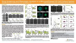 科学海报Drug Screening and Phenotypic Analysis in a Microwell-based 3D Cell Culture System
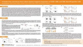 科学海报A Feeder-Free and Serum-Free Culture System Supports Expansion of CD34+ AML and CML Stem/Progenitor Cells
 沪公网安备31010102008431号