Zhang Y et al. ( 2018)
Nature communications 9 1 6
Nanoparticle anchoring targets immune agonists to tumors enabling anti-cancer immunity without systemic toxicity.
Immunostimulatory agents such as agonistic anti-CD137 and interleukin (IL)-2 generate effective anti-tumor immunity but also elicit serious toxicities,hampering their clinical application. Here we show that combination therapy with anti-CD137 and an IL-2-Fc fusion achieves significant initial anti-tumor activity,but also lethal immunotoxicity deriving from stimulation of circulating leukocytes. To overcome this toxicity,we demonstrate that anchoring IL-2 and anti-CD137 on the surface of liposomes allows these immune agonists to rapidly accumulate in tumors while lowering systemic exposure. In multiple tumor models,immunoliposome delivery achieves anti-tumor activity equivalent to free IL-2/anti-CD137 but with the complete absence of systemic toxicity. Immunoliposomes stimulated tumor infiltration by cytotoxic lymphocytes,cytokine production,and granzyme expression,demonstrating equivalent immunostimulatory effects to the free drugs in the local tumor microenvironment. Thus,surface-anchored particle delivery may provide a general approach to exploit the potent stimulatory activity of immune agonists without debilitating systemic toxicities.
View Publication
产品号#:
19753
19753RF
产品名:
Tan Q et al. (JAN 2018)
JCI insight 3 1
Activation-induced cytidine deaminase deficiency accelerates autoimmune diabetes in NOD mice.
B cells play an important role in type 1 diabetes (T1D) development. However,the role of B cell activation-induced cytidine deaminase (AID) in diabetes development is not clear. We hypothesized that AID is important in the immunopathogenesis of T1D. To test this hypothesis,we generated AID-deficient (AID-/-) NOD mice. We found that AID-/-NOD mice developed accelerated T1D,with worse insulitis and high levels of anti-insulin autoantibody in the circulation. Interestingly,neither maternal IgG transferred through placenta,nor IgA transferred through milk affected the accelerated diabetes development. AID-/-NOD mice showed increased activation and proliferation of B and T cells. We found enhanced T-B cell interactions in AID-/-NOD mice,with increased T-bet and IFN-γ expression in CD4+ T cells in the presence of AID-/- B cells. Moreover,excessive lymphoid expansion was observed in AID-/-NOD mice. Importantly,antigen-specific BDC2.5 CD4+ T cells caused more rapid onset of diabetes when cotransferred with AID-/- B cells than when cotransferred with AID+/+ B cells. Thus,our study provides insights into the role of AID in T1D. Our data also suggest that AID is a negative regulator of immune tolerance and ablation of AID can lead to exacerbated islet autoimmunity and accelerated T1D development.
View Publication
产品号#:
19854
19854RF
产品名:
EasySep™小鼠B细胞分选试剂盒
RoboSep™ 小鼠B细胞分选试剂盒
Wu X et al. (JAN 2018)
Cell 172 3 423--438.e25
Intrinsic Immunity Shapes Viral Resistance of Stem Cells.
Stem cells are highly resistant to viral infection compared to their differentiated progeny; however,the mechanism is mysterious. Here,we analyzed gene expression in mammalian stem cells and cells at various stages of differentiation. We find that,conserved across species,stem cells express a subset of genes previously classified as interferon (IFN) stimulated genes (ISGs) but that expression is intrinsic,as stem cells are refractory to interferon. This intrinsic ISG expression varies in a cell-type-specific manner,and many ISGs decrease upon differentiation,at which time cells become IFN responsive,allowing induction of a broad spectrum of ISGs by IFN signaling. Importantly,we show that intrinsically expressed ISGs protect stem cells against viral infection. We demonstrate the in vivo importance of intrinsic ISG expression for protecting stem cells and their differentiation potential during viral infection. These findings have intriguing implications for understanding stem cell biology and the evolution of pathogen resistance.
View Publication
产品号#:
02691
04434
04444
05110
05711
05712
05872
05873
18056
18056RF
72052
72054
72302
72304
72307
72308
70039
70039.1
70039.2
70039.3
70039.4
70039.5
70039.6
60062
60062BT
60062FI
60062FI.1
60062PE
60062PE.1
60045
60045AZ
60045AZ.1
60045BT
60045FI
60045FI.1
600
产品名:
StemSpan™ CD34+扩增添加物 (10X)
MethoCult™ H4434 Classic
MethoCult™ H4434 Classic
STEMdiff™定型内胚层检测试剂盒
NeuroCult™ SM1 神经添加物
CHIR99021
CHIR99021
Y-27632(二盐酸盐)
Y-27632(二盐酸盐)
Y-27632(二盐酸盐)
Y-27632(二盐酸盐)
抗人SSEA-4抗体,克隆号MC-813-70,生物素
抗人SSEA-4抗体,克隆号MC-813-70,FITC
抗人SSEA-4抗体, 克隆号MC-813-70,FITC
抗人SSEA-4抗体,克隆号MC-813-70,PE
抗人SSEA-4抗体,克隆号MC-813-70,PE
抗人CD90抗体,克隆5E10
抗人CD90抗体,克隆5E10,APC
抗人CD90抗体,克隆5E10,APC
抗人CD90抗体,克隆5E10,Biotin
抗人CD90抗体,克隆5E10,FITC
抗人CD90抗体,克隆5E10,FITC
Xu MM et al. (AUG 2017)
Immunity 47 2 363--373.e5
Dendritic Cells but Not Macrophages Sense Tumor Mitochondrial DNA for Cross-priming through Signal Regulatory Protein α Signaling.
Inhibition of cytosolic DNA sensing represents a strategy that tumor cells use for immune evasion,but the underlying mechanisms are unclear. Here we have shown that CD47-signal regulatory protein α (SIRPα) axis dictates the fate of ingested DNA in DCs for immune evasion. Although macrophages were more potent in uptaking tumor DNA,increase of DNA sensing by blocking the interaction of SIRPα with CD47 preferentially occurred in dendritic cells (DCs) but not in macrophages. Mechanistically,CD47 blockade enabled the activation of NADPH oxidase NOX2 in DCs,which in turn inhibited phagosomal acidification and reduced the degradation of tumor mitochondrial DNA (mtDNA) in DCs. mtDNA was recognized by cyclic-GMP-AMP synthase (cGAS) in the DC cytosol,contributing to type I interferon (IFN) production and antitumor adaptive immunity. Thus,our findings have demonstrated how tumor cells inhibit innate sensing in DCs and suggested that the CD47-SIRPα axis is critical for DC-driven antitumor immunity.
View Publication
产品号#:
18780
18780RF
18781
18781RF
19853
19853RF
70025
70025.1
70025.2
70025.3
产品名:
EasySep™小鼠CD11c正选试剂盒II
RoboSep™ 小鼠CD11c正选试剂盒II
EasySep™小鼠CD11c正选试剂盒II及脾脏解离液
RoboSep™ 小鼠CD11c正选试剂盒II及脾脏解离液
EasySep™小鼠CD8+ T细胞分选试剂盒
RoboSep™ 小鼠CD8+ T细胞分选试剂盒
冻存的人外周血单个核细胞
冻存的人外周血单个核细胞
冻存的人外周血单个核细胞
冻存的人外周血单个核细胞
Pekalski ML et al. (AUG 2017)
JCI insight 2 16
Neonatal and adult recent thymic emigrants produce IL-8 and express complement receptors CR1 and CR2.
The maintenance of peripheral naive T lymphocytes in humans is dependent on their homeostatic division,not continuing emigration from the thymus,which undergoes involution with age. However,postthymic maintenance of naive T cells is still poorly understood. Previously we reported that recent thymic emigrants (RTEs) are contained in CD31+CD25- naive T cells as defined by their levels of signal joint T cell receptor rearrangement excision circles (sjTRECs). Here,by differential gene expression analysis followed by protein expression and functional studies,we define that the naive T cells having divided the least since thymic emigration express complement receptors (CR1 and CR2) known to bind complement C3b- and C3d-decorated microbial products and,following activation,produce IL-8 (CXCL8),a major chemoattractant for neutrophils in bacterial defense. We also observed an IL-8-producing memory T cell subpopulation coexpressing CR1 and CR2 and with a gene expression signature resembling that of RTEs. The functions of CR1 and CR2 on T cells remain to be determined,but we note that CR2 is the receptor for Epstein-Barr virus,which is a cause of T cell lymphomas and a candidate environmental factor in autoimmune disease.
View Publication
产品号#:
15022
15062
产品名:
RosetteSep™人CD4+ T细胞富集抗体混合物
RosetteSep™人CD4+ T细胞富集抗体混合物
Chang C-F et al. (DEC 2017)
The Journal of clinical investigation
Erythrocyte efferocytosis modulates macrophages towards recovery after intracerebral hemorrhage.
Macrophages are a source of both proinflammatory and restorative functions in damaged tissue through complex dynamic phenotypic changes. Here,we sought to determine whether monocyte-derived macrophages (MDMs) contribute to recovery after acute sterile brain injury. By profiling the transcriptional dynamics of MDMs in the murine brain after experimental intracerebral hemorrhage (ICH),we found robust phenotypic changes in the infiltrating MDMs over time and demonstrated that MDMs are essential for optimal hematoma clearance and neurological recovery. Next,we identified the mechanism by which the engulfment of erythrocytes with exposed phosphatidylserine directly modulated the phenotype of both murine and human MDMs. In mice,loss of receptor tyrosine kinases AXL and MERTK reduced efferocytosis of eryptotic erythrocytes and hematoma clearance,worsened neurological recovery,exacerbated iron deposition,and decreased alternative activation of macrophages after ICH. Patients with higher circulating soluble AXL had poor 1-year outcomes after ICH onset,suggesting that therapeutically augmenting efferocytosis may improve functional outcomes by both reducing tissue injury and promoting the development of reparative macrophage responses. Thus,our results identify the efferocytosis of eryptotic erythrocytes through AXL/MERTK as a critical mechanism modulating macrophage phenotype and contributing to recovery from ICH.
View Publication

 EasySep™小鼠TIL(CD45)正选试剂盒


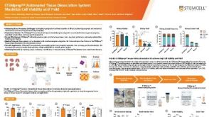 科学海报STEMprep™ Automated Tissue Dissociation System: Maximize Cell Viability and Yield
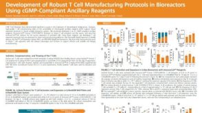 科学海报Development of Robust T Cell Manufacturing Protocols in Bioreactors Using cGMP-Compliant Ancillary Reagents


 沪公网安备31010102008431号