Chen H et al. (DEC 2015)
Biological research 48 1 59
Functional disruption of human leukocyte antigen II in human embryonic stem cell.
BACKGROUND Theoretically human embryonic stem cells (hESCs) have the capacity to self-renew and differentiate into all human cell types. Therefore,the greatest promise of hESCs-based therapy is to replace the damaged tissues of patients suffering from traumatic or degenerative diseases by the exact same type of cells derived from hESCs. Allograft immune rejection is one of the obstacles for hESCs-based clinical applications. Human leukocyte antigen (HLA) II leads to CD4(+) T cells-mediated allograft rejection. Hence,we focus on optimizing hESCs for clinic application through gene modification. RESULTS Transcription activator-like effector nucleases (TALENs) were used to target MHC class II transactivator (CIITA) in hESCs efficiently. CIITA (-/-) hESCs did not show any difference in the differentiation potential and self-renewal capacity. Dendritic cells (DCs) derived from CIITA (-/-) hESCs expressed CD83 and CD86 but without the constitutive HLA II. Fibroblasts derived from CIITA (-/-) hESCs were powerless in IFN-$\$ expression of HLA II. CONCLUSION We generated HLA II defected hESCs via deleting CIITA,a master regulator of constitutive and IFN-$\$ expression of HLA II genes. CIITA (-/-) hESCs can differentiate into tissue cells with non-HLA II expression. It's promising that CIITA (-/-) hESCs-derived cells could be used in cell therapy (e.g.,T cells and DCs) and escape the attack of receptors' CD4(+) T cells,which are the main effector cells of cellular immunity in allograft.
View Publication
产品号#:
05850
05857
05870
05875
85850
85857
85870
85875
产品名:
mTeSR™1
mTeSR™1
Shahbazi M et al. (JUL 2013)
Journal of the Neurological Sciences 330 1–2 85--93
Inhibitory effects of neural stem cells derived from human embryonic stem cells on differentiation and function of monocyte-derived dendritic cells
Neural stem cells (NSCs) possess immunosuppressive characteristics,but effects of NSCs on human dendritic cells (DCs),the most important antigen presenting cells,are less well studied. We used an in vitro approach to evaluate the effects of human NSCs on differentiation of human blood CD14+ monocytes into DCs. NSCs derived from H1 human embryonic stem cells (hESC-NSCs) and human ReNcell NSC line,as well as human bone marrow derived mesenchymal stem cells (MSCs),were tested. We observed that in response to treatment with interleukin-4 and granulocyte macrophage colony-stimulating factor CD14+ monocytes co-cultured with NSCs were able to down-regulate CD14 and up-regulate the differentiation marker CD1a,whereas MSC co-culture strongly inhibited CD1a expression and supported prolonged expression of CD14. A similar difference between NSCs and MSCs was noted when lipopolysaccharides were included to induce maturation of monocyte-derived DCs. However,when effects on the function of derived DCs were investigated,NSCs suppressed the elevation of the DC maturation marker CD83,although not the up-regulation of costimulatory molecules CD80,CD86 and CD40,and impaired the functional capacity of the derived DCs to stimulate alloreactive T cells. We did not observe any obvious difference between hESC-NSCs and ReNcell NSCs in inhibiting DC maturation and function. Our data suggest that although human NSCs are less effective than human MSCs in suppressing monocyte differentiation into DCs,these stem cells can still affect the function of DCs,ultimately regulating specific immune responses.
View Publication
Zeng J et al. (MAY 2012)
The Journal of Immunology 188 9 4297--4304
Enhancing Immunostimulatory Function of Human Embryonic Stem Cell-Derived Dendritic Cells by CD1d Overexpression
Human embryonic stem cell-derived dendritic cells (hESC-DCs) may potentially provide a platform to generate off-the-shelf" therapeutic cancer vaccines. To apply hESC-DCs for cancer immunotherapy in a semiallogeneic setting�
View Publication
Begum AN et al. (JUL 2014)
Translational psychiatry 4 January e414
Women with the Alzheimer's risk marker ApoE4 lose A-specific CD4 T cells 10-20 years before men.
Adaptive immunity to self-antigens causes autoimmune disorders,such as multiple sclerosis,psoriasis and type 1 diabetes; paradoxically,T- and B-cell responses to amyloid-$\$(A$\$) reduce Alzheimer's disease (AD)-associated pathology and cognitive impairment in mouse models of the disease. The manipulation of adaptive immunity has been a promising therapeutic approach for the treatment of AD,although vaccine and anti-A$\$ approaches have proven difficult in patients,thus far. CD4(+) T cells have a central role in regulating adaptive immune responses to antigens,and A$\$-specific CD4(+) T cells have been shown to reduce AD pathology in mouse models. As these cells may facilitate endogenous mechanisms that counter AD,an evaluation of their abundance before and during AD could provide important insights. A$\$-CD4see is a new assay developed to quantify A$\$-specific CD4(+) T cells in human blood,using dendritic cells derived from human pluripotent stem cells. In tests of textgreater50 human subjects A$\$-CD4see showed an age-dependent decline of A$\$-specific CD4(+) T cells,which occurs earlier in women than men. In aggregate,men showed a 50% decline in these cells by the age of 70 years,but women reached the same level before the age of 60 years. Notably,women who carried the AD risk marker apolipoproteinE-ɛ4 (ApoE4) showed the earliest decline,with a precipitous drop between 45 and 52 years,when menopause typically begins. A$\$-CD4see requires a standard blood draw and provides a minimally invasive approach for assessing changes in A$\$ that may reveal AD-related changes in physiology by a decade. Furthermore,CD4see probes can be modified to target any peptide,providing a powerful new tool to isolate antigen-specific CD4(+) T cells from human subjects.
View Publication


 EasySep™小鼠TIL(CD45)正选试剂盒




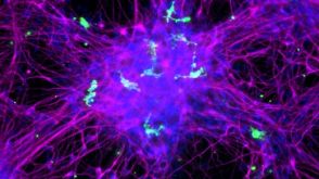 实验方案How to Co-Culture Human Pluripotent Stem Cell (hPSC)-Derived Forebrain Neurons and Microglia
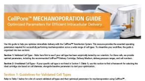
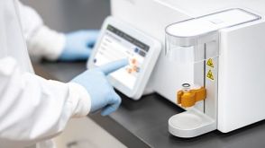 实验方案Gene Editing Human Pluripotent Stem Cells (hPSCs) Using the CellPore™ Transfection System
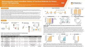 科学海报Mechanoporation-Based Intracellular Delivery of Functional Molecules for Primary Immune Cell Engineering


 沪公网安备31010102008431号