Osman MS et al. (FEB 2007)
Journal of immunology (Baltimore,Md. : 1950) 178 3 1261--7
Activating Ly-49 receptors regulate LFA-1-mediated adhesion by NK cells.
NK cells are important for innate resistance to tumors and viruses. Engagement of activating Ly-49 receptors expressed by NK cells leads to rapid NK cell activation resulting in target cell lysis and cytokine production. The ITAM-containing DAP12 adapter protein stably associates with activating Ly-49 receptors,and couples receptor recognition with generation of NK responses. Activating Ly-49s are potent stimulators of murine NK cell functions,yet how they mediate such activities is not well understood. We demonstrate that these receptors trigger LFA-1-dependent tight conjugation between NK cells and target cells. Furthermore,we show that activating Ly-49 receptor engagement leads to rapid DAP12-dependent up-regulation of NK cell LFA-1 adhesiveness to ICAM-1 that is also dependent on tyrosine kinases of the Syk and Src families. These results indicate for the first time that activating Ly-49s control adhesive properties of LFA-1,and by DAP12-dependent inside-out signaling. Ly-49-driven mobilization of LFA-1 adhesive function may represent a fundamental proximal event during NK cell interactions with target cells involving activating Ly-49 receptors,leading to target cell death.
View Publication
产品号#:
18755
18755RF
产品名:
EasySep™小鼠CD49b正选试剂盒
RoboSep™ 小鼠CD49b正选试剂盒含滤芯吸头
Tang Y et al. (SEP 2007)
Journal of immunology (Baltimore,Md. : 1950) 179 5 2815--23
Regulation of antibody-dependent cellular cytotoxicity by IgG intrinsic and apparent affinity for target antigen.
Unconjugated mAbs have emerged as useful cancer therapeutics. Ab-dependent cellular cytotoxicity (ADCC) is believed to be a major antitumor mechanism of some anticancer Abs. However,the factors that regulate the magnitude of ADCC are incompletely understood. In this study,we described the relationship between Ab affinity and ADCC. A series of human IgG1 isotype Abs was created from the anti-HER2/neu (also named c-erbB2) C6.5 single-chain Fv (scFv) and its affinity mutants. The scFv affinities range from 10(-7) to 10(-11) M,and the IgG Abs retain the affinities of the scFv from which they were derived. The apparent affinity of the Abs ranged from nearly 10(-10) M (the lowest affinity variant) to almost 10(-11) M (the other variants). The IgG molecules were tested for their ability to elicit ADCC in vitro against three tumor cell lines with differing levels of HER2/neu expression using unactivated human PBMC from healthy donors as the effector cells. The results demonstrated that both the apparent affinity and intrinsic affinity of the Abs studied regulate ADCC. High-affinity tumor Ag binding by the IgGs led to the most efficient and powerful ADCC. Tumor cells expressing high levels of HER2/neu are more susceptible to the ADCC triggered by Abs than the cells expressing lower amounts of HER2/neu. These findings justify the examination of high affinity Abs for ADCC promotion. Because high affinity may impair in vivo tumor targeting,a careful examination of Ab structure to function relationships is required to develop optimized therapeutic unconjugated Abs.
View Publication
产品号#:
15025
15065
产品名:
RosetteSep™人NK细胞富集抗体混合物
RosetteSep™人NK细胞富集抗体混合物
Norman JM et al. (OCT 2011)
Nature immunology 12 10 975--83
The antiviral factor APOBEC3G enhances the recognition of HIV-infected primary T cells by natural killer cells.
APOBEC3G (A3G) is an intrinsic antiviral factor that inhibits the replication of human immunodeficiency virus (HIV) by deaminating cytidine residues to uridine. This causes guanosine-to-adenosine hypermutation in the opposite strand and results in inactivation of the virus. HIV counteracts A3G through the activity of viral infectivity factor (Vif),which promotes degradation of A3G. We report that viral protein R (Vpr),which interacts with a uracil glycosylase,also counteracted A3G by diminishing the incorporation of uridine. However,this process resulted in activation of the DNA-damage–response pathway and the expression of natural killer (NK) cell–activating ligands. Our results show that pathogen-induced deamination of cytidine and the DNA-damage response to virus-mediated repair of the incorporation of uridine enhance the recognition of HIV-infected cells by NK cells.
View Publication
产品号#:
17755
产品名:
Garg TK et al. (SEP 2012)
Haematologica 97 9 1348--56
Highly activated and expanded natural killer cells for multiple myeloma immunotherapy.
BACKGROUND Patients with gene expression profiling-defined high-risk myeloma in relapse have poor outcomes with current therapies. We tested whether natural killer cells expanded by co-culture with K562 cells transfected with 41BBL and membrane-bound interleukin-15 could kill myeloma cells with a high-risk gene expression profile in vitro and in a unique model which recapitulates human myeloma. DESIGN AND METHODS OPM2 and high-risk primary myeloma tumors were grown in human fetal bone implanted into non-obese diabetic severe combined immunodeficiency mice with a deficient interleukin-2 receptor gamma chain. These mice are devoid of endogenous natural killer and T-cell activity and were used to determine whether adoptively transferred expanded natural killer cells could inhibit myeloma growth and myeloma-associated bone destruction. RESULTS Natural killer cells from healthy donors and myeloma patients expanded a median of 804- and 351-fold,respectively,without significant T-cell expansion. Expanded natural killer cells killed both allogeneic and autologous primary myeloma cells avidly via a perforin-mediated mechanism in which the activating receptor NKG2D,natural cytotoxicity receptors,and DNAX-accessory molecule-1 played a central role. Adoptive transfer of expanded natural killer cells inhibited the growth of established OPM2 and high-risk primary myeloma tumors grown in the murine model. The transferred,expanded natural killer cells proliferated in vivo in an interleukin-2 dose-dependent fashion,persisted up to 4 weeks,were readily detectable in the human bone,inhibited myeloma growth and protected bone from myeloma-induced osteolysis. CONCLUSIONS These studies provide the rationale for testing expanded natural killer cells in humans.
View Publication
产品号#:
19055
19055RF
产品名:
EasySep™人NK细胞富集试剂盒
RoboSep™ 人NK细胞富集试剂盒含滤芯吸头
Kang L et al. ( 2013)
Frontiers in immunology 4 MAY 101
Characterization and ex vivo Expansion of Human Placenta-Derived Natural Killer Cells for Cancer Immunotherapy.
Recent clinical studies suggest that adoptive transfer of donor-derived natural killer (NK) cells may improve clinical outcome in hematological malignancies and some solid tumors by direct anti-tumor effects as well as by reduction of graft versus host disease (GVHD). NK cells have also been shown to enhance transplant engraftment during allogeneic hematopoietic stem cell transplantation (HSCT) for hematological malignancies. The limited ex vivo expansion potential of NK cells from peripheral blood (PB) or umbilical cord blood (UCB) has however restricted their therapeutic potential. Here we define methods to efficiently generate NK cells from donor-matched,full-term human placenta perfusate (termed Human Placenta-Derived Stem Cell,HPDSC) and UCB. Following isolation from cryopreserved donor-matched HPDSC and UCB units,CD56+CD3- placenta-derived NK cells,termed pNK cells,were expanded in culture for up to 3 weeks to yield an average of 1.2 billion cells per donor that were textgreater80% CD56+CD3-,comparable to doses previously utilized in clinical applications. Ex vivo-expanded pNK cells exhibited a marked increase in anti-tumor cytolytic activity coinciding with the significantly increased expression of NKG2D,NKp46,and NKp44 (p textless 0.001,p textless 0.001,and p textless 0.05,respectively). Strong cytolytic activity was observed against a wide range of tumor cell lines in vitro. pNK cells display a distinct microRNA (miRNA) expression profile,immunophenotype,and greater anti-tumor capacity in vitro compared to PB NK cells used in recent clinical trials. With further development,pNK may represent a novel and effective cellular immunotherapy for patients with high clinical needs and few other therapeutic options.
View Publication
E. Giuliani et al. (mar 2019)
Scientific reports 9 1 4373
Hexamethylene bisacetamide impairs NK cell-mediated clearance of acute T lymphoblastic leukemia cells and HIV-1-infected T cells that exit viral latency.
The hexamethylene bisacetamide (HMBA) anticancer drug was dismissed due to limited efficacy in leukemic patients but it may re-enter into the clinics in HIV-1 eradication strategies because of its recently disclosed capacity to reactivate latent virus. Here,we investigated the impact of HMBA on the cytotoxicity of natural killer (NK) cells against acute T lymphoblastic leukemia (T-ALL) cells or HIV-1-infected T cells that exit from latency. We show that in T-ALL cells HMBA upmodulated MICB and ULBP2 ligands for the NKG2D activating receptor. In a primary CD4+ T cell-based latency model,HMBA did not reactivate HIV-1,yet enhanced ULBP2 expression on cells harboring virus reactivated by prostratin (PRO). However,HMBA reduced the expression of NKG2D and its DAP10 adaptor in NK cells,hence impairing NKG2D-mediated cytotoxicity and DAP10-dependent response to IL-15 stimulation. Alongside,HMBA dampened killing of T-ALL targets by IL-15-activated NK cells and impaired NK cell-mediated clearance of PRO-reactivated HIV-1+ cells. Overall,our results demonstrate a dominant detrimental effect of HMBA on the NKG2D pathway that crucially controls NK cell-mediated killing of tumors and virus-infected cells,providing one possible explanation for poor clinical outcome in HMBA-treated cancer patients and raising concerns for future therapeutic application of this drug.
View Publication
产品号#:
19052
19052RF
产品名:
EasySep™人CD4+ T细胞富集试剂盒
RoboSep™ 人CD4+ T细胞富集试剂盒含滤芯吸头
A. Lisco et al. (apr 2019)
JCI insight 4 8
Identification of rare HIV-1-infected patients with extreme CD4+ T cell decline despite ART-mediated viral suppression.
BACKGROUND The goal of antiretroviral therapy (ART) is to suppress HIV-1 replication and reconstitute CD4+ T cells. Here,we report on HIV-infected individuals who had a paradoxical decline in CD4+ T cells despite ART-mediated suppression of plasma HIV-1 load (pVL). We defined such an immunological outcome as extreme immune decline (EXID). METHODS EXID's clinical and immunological characteristics were compared to immunological responders (IRs),immunological nonresponders (INRs),healthy controls (HCs),and idiopathic CD4+ lymphopenia (ICL) patients. T cell immunophenotyping and assembly/activation of inflammasomes were evaluated by flow cytometry. PBMC transcriptome analysis and genetic screening for pathogenic variants were performed. Levels of cytokines/chemokines were measured by electrochemiluminescence. Luciferase immunoprecipitation system and NK-mediated antibody-dependent cellular cytotoxicity (ADCC) assays were used to identify anti-lymphocyte autoantibodies. RESULTS EXIDs were infected with non-B HIV-1 subtypes and after 192 weeks of consistent ART-mediated pVL suppression had a median CD4+ decrease of 157 cells/mul,compared with CD4+ increases of 193 cells/mul and 427 cells/mul in INR and IR,respectively. EXID had reduced naive CD4+ T cells,but similar proportions of cycling CD4+ T cells and HLA-DR+CD38+CD8+ T cells compared with IR and INR. Levels of inflammatory cytokines were also similar in EXID and INR,but the IL-7 axis was profoundly perturbed compared with HC,IR,INR,and ICL. Genes involved in T cell and monocyte/macrophage function,autophagy,and cell migration were differentially expressed in EXID. Two of the 5 EXIDs had autoantibodies causing ADCC,while 2 different EXIDs had an increased inflammasome/caspase-1 activation despite consistently ART-suppressed pVL. CONCLUSIONS EXID is a distinct immunological outcome compared with previously described INR. Anti-CD4+ T cell autoantibodies and aberrant inflammasome/caspase-1 activation despite suppressed HIV-1 viremia are among the mechanisms responsible for EXID.
View Publication
产品号#:
17955
17955RF
100-0960
产品名:
EasySep™人NK细胞分选试剂盒
RoboSep™ 人NK细胞分选试剂盒
EasySep™人NK细胞分离试剂盒
T. A. Mace et al. (mar 2019)
Scientific reports 9 1 5068
Soy isoflavones and their metabolites modulate cytokine-induced natural killer cell function.
Soybeans are a rich source of isoflavones that have been linked with anti-inflammatory processes and various health benefits. However,specific mechanisms whereby soy bioactives impact immune cell subsets are unclear. Isoflavones,such as genistein and daidzein,are metabolized by microbes to bioactive metabolites as O-desmethylangolensin (O-DMA) and equol,whose presence has been linked to health benefits. We examined how soy isoflavones and metabolites impact natural killer (NK) cell signaling and function. We observe no impact of isoflavones on viability of healthy donor peripheral blood mononuclear cells (PBMCs) or NK cells,even at high (25 µM) concentrations. However,pre-treatment of PBMCs with physiologically-relevant concentrations of genistein (p = 0.0023) and equol (p = 0.006) decreases interleukin (IL)-12/IL-18-induced interferon-gamma (IFN-gamma) production versus controls. Detailed cellular analyses indicate genistein and equol decrease IL-12/IL-18-induced IFN-gamma production by human NK cell subsets,but do not consistently alter cytotoxicity. At the level of signal transduction,genistein decreases IL-12/IL-18-induced total phosphorylated tyrosine,and phosphorylation MAPK pathway components. Further,genistein limits IL-12/IL-18-mediated upregulation of IL-18Ralpha expression on NK cells (p = 0.0109). Finally,in vivo studies revealed that C57BL/6 mice fed a soy-enriched diet produce less plasma IFN-gamma following administration of IL-12/IL-18 versus control-fed animals (p {\textless} 0.0001). This study provides insight into how dietary soy modulates NK cell functions.
View Publication


 EasySep™小鼠TIL(CD45)正选试剂盒




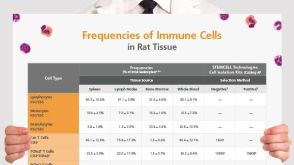 挂图Frequencies of Immune Cells in Rat Tissue Lists the estimated frequencies of more than 15 immune cell types in Sprague Dawley rats

 沪公网安备31010102008431号