Le MX et al. (NOV 2016)
Scientific reports 6 37215
Kin17 facilitates multiple double-strand break repair pathways that govern B cell class switching.
Class switch recombination (CSR) in B cells requires the timely repair of DNA double-stranded breaks (DSBs) that result from lesions produced by activation-induced cytidine deaminase (AID). Through a genome-wide RNAi screen,we identified Kin17 as a gene potentially involved in the maintenance of CSR in murine B cells. In this study,we confirm a critical role for Kin17 in CSR independent of AID activity. Furthermore,we make evident that DSBs generated by AID or ionizing radiation require Kin17 for efficient repair and resolution. Our report shows that reduced Kin17 results in an elevated deletion frequency following AID mutational activity in the switch region. In addition,deficiency in Kin17 affects the functionality of multiple DSB repair pathways,namely homologous recombination,non-homologous end-joining,and alternative end-joining. This report demonstrates the importance of Kin17 as a critical factor that acts prior to the repair phase of DSB repair and is of bona fide importance for CSR.
View Publication
产品号#:
19854
19854RF
产品名:
EasySep™小鼠B细胞分选试剂盒
RoboSep™ 小鼠B细胞分选试剂盒
Li MMH et al. (NOV 2016)
The Journal of experimental medicine
Interferon regulatory factor 2 protects mice from lethal viral neuroinvasion.
The host responds to virus infection by activating type I interferon (IFN) signaling leading to expression of IFN-stimulated genes (ISGs). Dysregulation of the IFN response results in inflammatory diseases and chronic infections. In this study,we demonstrate that IFN regulatory factor 2 (IRF2),an ISG and a negative regulator of IFN signaling,influences alphavirus neuroinvasion and pathogenesis. A Sindbis virus strain that in wild-type (WT) mice only causes disease when injected into the brain leads to lethal encephalitis in Irf2(-/-) mice after peripheral inoculation. Irf2(-/-) mice fail to control virus replication and recruit immune infiltrates into the brain. Reduced B cells and virus-specific IgG are observed in the Irf2(-/-) mouse brains despite the presence of peripheral neutralizing antibodies,suggesting a defect in B cell trafficking to the central nervous system (CNS). B cell-deficient μMT mice are significantly more susceptible to viral infection,yet WT B cells and serum are unable to rescue the Irf2(-/-) mice. Collectively,our data demonstrate that proper localization of B cells and local production of antibodies in the CNS are required for protection. The work advances our understanding of host mechanisms that affect viral neuroinvasion and their contribution to immunity against CNS infections.
View Publication
产品号#:
19854
19854RF
产品名:
EasySep™小鼠B细胞分选试剂盒
RoboSep™ 小鼠B细胞分选试剂盒
Inoue S et al. (AUG 2006)
Cancer research 66 15 7741--7
Inhibitory effects of B cells on antitumor immunity.
B-cell functions in antitumor immunity are not well understood. In this study,we evaluated the role of B cells in the development of antitumor immunity using Friend murine leukemia virus gag-expressing mouse EL-4 (EL-4 gag),D5 mouse melanoma,or MCA304 mouse sarcoma cells. To screen tumors for susceptibility to B-cell-deficient immune environments,spleen cells from naive C57BL/6 [wild-type (WT)] and B-cell knockout (BKO) mice were cultured with irradiated tumor cells in vitro. When cells were stimulated with EL-4 gag or D5 (but not MCA304 tumors),IFN-gamma production from CD8 T cells and natural killer cells was markedly decreased in WT compared with BKO cultures. IFN-gamma production was correlated with CD40 ligand expression on the tumor and inversely with interleukin-10 (IL-10) production by B cells. Sorted WT B cells produced more IL-10 than CD40 knockout (CD40KO) B cells when cocultured with EL-4 gag or D5 (but not MCA304). IFN-gamma production by BKO cells was reduced by the addition of sorted naive WT B cells (partially by CD40KO B cells) or recombinant mouse IL-10. In vivo tumor progression mirrored in vitro studies in that WT mice were unable to control tumor growth whereas EL-4 gag and D5 tumors (but not MCA304) were eliminated in BKO mice. Robust in vivo antitumor CTLs developed only in BKO tumor-challenged mice. Our studies provide the first mechanistic basis for the concept that B-cell depletion could therapeutically enhance antitumor immune responses to certain tumors by decreasing IL-10 production from B cells.
View Publication

 EasySep™小鼠TIL(CD45)正选试剂盒


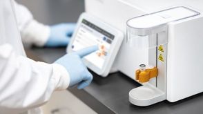 实验方案Optimizing Delivery Efficiency with Fluorescent Dextran Using the CellPore™ Transfection System


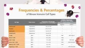 挂图Frequencies and Percentages of Mouse Immune Cell Types List of the frequencies of over 25 immune cell types in C57BL/6 mice
 沪公网安备31010102008431号