de Valle E et al. (APR 2016)
The Journal of Experimental Medicine 213 4 621--41
NFκB1 is essential to prevent the development of multiorgan autoimmunity by limiting IL-6 production in follicular B cells.
We examined the role of NFκB1 in the homeostasis and function of peripheral follicular (Fo) B cells. Aging mice lacking NFκB1 (Nfκb1(-/-)) develop lymphoproliferative and multiorgan autoimmune disease attributed in large part to the deregulated activity ofNfκb1(-/-)Fo B cells that produce excessive levels of the proinflammatory cytokine interleukin 6 (IL-6). Despite enhanced germinal center (GC) B cell differentiation,the formation of GC structures was severely disrupted in theNfκb1(-/-)mice. Bone marrow chimeric mice revealed that the Fo B cell-intrinsic loss of NFκB1 led to the spontaneous generation of GC B cells. This was primarily the result of an increase in IL-6 levels,which promotes the differentiation of Fo helper CD4(+)T cells and acts in an autocrine manner to reduce antigen receptor and toll-like receptor activation thresholds in a population of proliferating IgM(+)Nfκb1(-/-)Fo B cells. We demonstrate that p50-NFκB1 repressesIl-6transcription in Fo B cells,with the loss of NFκB1 also resulting in the uncontrolled RELA-driven transcription ofIl-6.Collectively,our findings identify a previously unrecognized role for NFκB1 in preventing multiorgan autoimmunity through its negative regulation ofIl-6gene expression in Fo B cells.
View Publication
产品号#:
19854
19854RF
产品名:
EasySep™小鼠B细胞分选试剂盒
RoboSep™ 小鼠B细胞分选试剂盒
Flach A-C et al. (MAR 2016)
Proceedings of the National Academy of Sciences of the United States of America 113 12 3323--8
Autoantibody-boosted T-cell reactivation in the target organ triggers manifestation of autoimmune CNS disease.
Multiple sclerosis (MS) is caused by T cells that are reactive for brain antigens. In experimental autoimmune encephalomyelitis,the animal model for MS,myelin-reactive T cells initiate the autoimmune process when entering the nervous tissue and become reactivated upon local encounter of their cognate CNS antigen. Thereby,the strength of the T-cellular reactivation process within the CNS tissue is crucial for the manifestation and the severity of the clinical disease. Recently,B cells were found to participate in the pathogenesis of CNS autoimmunity,with several diverse underlying mechanisms being under discussion. We here report that B cells play an important role in promoting the initiation process of CNS autoimmunity. Myelin-specific antibodies produced by autoreactive B cells after activation in the periphery diffused into the CNS together with the first invading pathogenic T cells. The antibodies accumulated in resident antigen-presenting phagocytes and significantly enhanced the activation of the incoming effector T cells. The ensuing strong blood-brain barrier disruption and immune cell recruitment resulted in rapid manifestation of clinical disease. Therefore,myelin oligodendrocyte glycoprotein (MOG)-specific autoantibodies can initiate disease bouts by cooperating with the autoreactive T cells in helping them to recognize their autoantigen and become efficiently reactivated within the immune-deprived nervous tissue.
View Publication
产品号#:
19851
19851RF
19854
19854RF
产品名:
EasySep™小鼠T细胞分选试剂盒
RoboSep™ 小鼠T细胞分选试剂盒
EasySep™小鼠B细胞分选试剂盒
RoboSep™ 小鼠B细胞分选试剂盒
Thompson EA et al. (APR 2016)
Journal of Immunology 196 7 3054--63
Shortened Intervals during Heterologous Boosting Preserve Memory CD8 T Cell Function but Compromise Longevity.
Developing vaccine strategies to generate high numbers of Ag-specific CD8 T cells may be necessary for protection against recalcitrant pathogens. Heterologous prime-boost-boost immunization has been shown to result in large quantities of functional memory CD8 T cells with protective capacities and long-term stability. Completing the serial immunization steps for heterologous prime-boost-boost can be lengthy,leaving the host vulnerable for an extensive period of time during the vaccination process. We show in this study that shortening the intervals between boosting events to 2 wk results in high numbers of functional and protective Ag-specific CD8 T cells. This protection is comparable to that achieved with long-term boosting intervals. Short-boosted Ag-specific CD8 T cells display a canonical memory T cell signature associated with long-lived memory and have identical proliferative potential to long-boosted T cells Both populations robustly respond to antigenic re-exposure. Despite this,short-boosted Ag-specific CD8 T cells continue to contract gradually over time,which correlates to metabolic differences between short- and long-boosted CD8 T cells at early memory time points. Our studies indicate that shortening the interval between boosts can yield abundant,functional Ag-specific CD8 T cells that are poised for immediate protection; however,this is at the expense of forming stable long-term memory.
View Publication
产品号#:
17951
17951RF
17952
17952RF
19254
19254RF
19853
19853RF
100-0695
100-0696
产品名:
EasySep™人T细胞分选试剂盒
RoboSep™ 人T细胞分选试剂盒
EasySep™人CD4+ T细胞分选试剂盒
RoboSep™ 人CD4+ T细胞分选试剂盒
EasySep™人Naïve B细胞富集试剂盒
RoboSep™ 人Naïve B细胞富集试剂盒含滤芯吸头
EasySep™小鼠CD8+ T细胞分选试剂盒
RoboSep™ 小鼠CD8+ T细胞分选试剂盒
EasySep™人T细胞分选试剂盒
EasySep™人CD4+ T细胞分离试剂盒
Tan Q et al. (JAN 2018)
JCI insight 3 1
Activation-induced cytidine deaminase deficiency accelerates autoimmune diabetes in NOD mice.
B cells play an important role in type 1 diabetes (T1D) development. However,the role of B cell activation-induced cytidine deaminase (AID) in diabetes development is not clear. We hypothesized that AID is important in the immunopathogenesis of T1D. To test this hypothesis,we generated AID-deficient (AID-/-) NOD mice. We found that AID-/-NOD mice developed accelerated T1D,with worse insulitis and high levels of anti-insulin autoantibody in the circulation. Interestingly,neither maternal IgG transferred through placenta,nor IgA transferred through milk affected the accelerated diabetes development. AID-/-NOD mice showed increased activation and proliferation of B and T cells. We found enhanced T-B cell interactions in AID-/-NOD mice,with increased T-bet and IFN-γ expression in CD4+ T cells in the presence of AID-/- B cells. Moreover,excessive lymphoid expansion was observed in AID-/-NOD mice. Importantly,antigen-specific BDC2.5 CD4+ T cells caused more rapid onset of diabetes when cotransferred with AID-/- B cells than when cotransferred with AID+/+ B cells. Thus,our study provides insights into the role of AID in T1D. Our data also suggest that AID is a negative regulator of immune tolerance and ablation of AID can lead to exacerbated islet autoimmunity and accelerated T1D development.
View Publication
Biologic and genetic characterization of the novel amyloidogenic lambda light chain-secreting human cell lines, ALMC-1 and ALMC-2.
Primary systemic amyloidosis (AL) is a rare monoclonal plasma cell (PC) disorder characterized by the deposition of misfolded immunoglobulin (Ig) light chains (LC) in vital organs throughout the body. To our knowledge,no cell lines have ever been established from AL patients. Here we describe the establishment of the ALMC-1 and ALMC-2 cell lines from an AL patient. Both cell lines exhibit a PC phenotype and display cytokine-dependent growth. Using a comprehensive genetic approach,we established the genetic relationship between the cell lines and the primary patient cells,and we were also able to identify new genetic changes accompanying tumor progression that may explain the natural history of this patient's disease. Importantly,we demonstrate that free lambda LC secreted by both cell lines contained a beta structure and formed amyloid fibrils. Despite absolute Ig LC variable gene sequence identity,the proteins show differences in amyloid formation kinetics that are abolished by the presence of Na(2)SO(4). The formation of amyloid fibrils from these naturally secreting human LC cell lines is unprecedented. Moreover,these cell lines will provide an invaluable tool to better understand AL,from the combined perspectives of amyloidogenic protein structure and amyloid formation,genetics,and cell biology.
View Publication
产品号#:
18357
18357RF
21000
20119
20155
18387
18387RF
产品名:
RoboSep™- S
RoboSep™ 吸头组件抛光剂
RoboSep™分选管套装(9个塑料管)
Douglas KB et al. (JUL 2009)
Genes and immunity 10 5 457--69
Complement receptor 2 polymorphisms associated with systemic lupus erythematosus modulate alternative splicing.
Genetic factors influence susceptibility to systemic lupus erythematosus (SLE). A recent family-based analysis in Caucasian and Chinese populations provided evidence for association of single-nucleotide polymorphisms (SNPs) in the complement receptor 2 (CR2/CD21) gene with SLE. Here we confirmed this result in a case-control analysis of an independent European-derived population including 2084 patients with SLE and 2853 healthy controls. A haplotype formed by the minor alleles of three CR2 SNPs (rs1048971,rs17615,rs4308977) showed significant association with decreased risk of SLE (30.4% in cases vs 32.6% in controls,P=0.016,OR=0.90 (0.82-0.98)). Two of these SNPs are in exon 10,directly 5' of an alternatively spliced exon preferentially expressed in follicular dendritic cells (FDC),and the third is in the alternatively spliced exon. Effects of these SNPs and a fourth SNP in exon 11 (rs17616) on alternative splicing were evaluated. We found that the minor alleles of these SNPs decreased splicing efficiency of exon 11 both in vitro and ex vivo. These findings further implicate CR2 in the pathogenesis of SLE and suggest that CR2 variants alter the maintenance of tolerance and autoantibody production in the secondary lymphoid tissues where B cells and FDCs interact.
View Publication

 EasySep™小鼠TIL(CD45)正选试剂盒


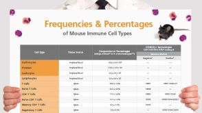 挂图Frequencies and Percentages of Mouse Immune Cell Types List of the frequencies of over 25 immune cell types in C57BL/6 mice

 沪公网安备31010102008431号