D. Park et al. (may 2019)
Scientific reports 9 1 7094
Differences in the molecular signatures of mucosal-associated invariant T cells and conventional T cells.
Mucosal-associated invariant T (MAIT) cells exhibit different characteristics from those of TCRalpha7.2- conventional T cells. They play important roles in various inflammatory diseases,including rheumatoid arthritis and inflammatory bowel disease. MAIT cells express a single T cell receptor alpha chain,TCRalpha7.2 segment associated with Jalpha33 and CDR3 with fixed length,which recognizes bacteria-derived vitamin B metabolites. However,the characteristics of MAIT cells and TCRalpha7.2+ CD161- T cells have never been compared. Here,we performed RNA sequencing to compare the properties of MAIT cells,TCRalpha7.2- conventional T cells and TCRalpha7.2+ CD161- T cells. Genome-wide transcriptomes of MAIT cells,TCRalpha7.2- conventional T cells,and TCRalpha7.2+ CD161- T cells were compared and analyzed using causal network analysis. This is the first report comparing the transcriptomes of MAIT cells,TCRalpha7.2- conventional T cells and TCRalpha7.2+ CD161- T cells. We also identified the predominant signaling pathways of MAIT cells,which differed from those of TCRalpha7.2- conventional T cells and TCRalpha7.2+ CD161- T cells,through a gene set enrichment test and upstream regulator analysis and identified the genes responsible for the characteristic MAIT cell phenotypes. Our study advances the complete understanding of MAIT biology.
View Publication
产品号#:
15021
15061
产品名:
RosetteSep™人T细胞富集抗体混合物
RosetteSep™人T细胞富集抗体混合物
D. M. Previte et al. (apr 2019)
Cell reports 27 1 129--141.e4
Lymphocyte Activation Gene-3 Maintains Mitochondrial and Metabolic Quiescence in Naive CD4+ T Cells.
Lymphocyte activation gene-3 (LAG-3) is an inhibitory receptor expressed by CD4+ T cells and tempers their homeostatic expansion. Because CD4+ T cell proliferation is tightly coupled to bioenergetics,we investigate the role of LAG-3 in modulating naive CD4+ T cell metabolism. LAG-3 deficiency enhances the metabolic profile of naive CD4+ T cells by elevating levels of mitochondrial biogenesis. In vivo,LAG-3 blockade partially restores expansion and the metabolic phenotype of wild-type CD4+ T cells to levels of Lag3-/- CD4+ T cells,solidifying that LAG-3 controls these processes. Lag3-/- CD4+ T cells also demonstrate greater signal transducer and activator of transcription 5 (STAT5) activation,enabling resistance to interleukin-7 (IL-7) deprivation. These results implicate this pathway as a target of LAG-3-mediated inhibition. Additionally,enhancement of STAT5 activation,as a result of LAG-3 deficiency,contributes to greater activation potential in these cells. These results identify an additional mode of regulation elicited by LAG-3 in controlling CD4+ T cell responses.
View Publication
产品号#:
19852
19852RF
产品名:
EasySep™小鼠CD4+ T细胞分选试剂盒
RoboSep™ 小鼠CD4+ T细胞分选试剂盒
Billard E et al. (OCT 2007)
Infection and immunity 75 10 4980--9
Brucella suis prevents human dendritic cell maturation and antigen presentation through regulation of tumor necrosis factor alpha secretion.
Brucella is a facultative intracellular pathogen and the etiological agent of brucellosis. In some cases,human brucellosis results in a persistent infection that may reactivate years after the initial exposure. The mechanisms by which the parasite evades clearance by the immune response to chronically infect its host are unknown. We recently demonstrated that dendritic cells (DCs),which are critical components of adaptive immunity,are highly susceptible to Brucella infection and are a preferential niche for the development of the bacteria. Here,we report that in contrast to several intracellular bacteria,Brucella prevented the infected DCs from engaging in their maturation process and impaired their capacities to present antigen to naïve T cells and to secrete interleukin-12. Moreover,Brucella-infected DCs failed to release tumor necrosis factor alpha (TNF-alpha),a defect involving the bacterial protein Omp25. Exogenous TNF-alpha addition to Brucella-infected DCs restored cell maturation and allowed them to present antigens. Two avirulent mutants of B. suis,B. suis bvrR and B. suis omp25 mutants,which do not express the Omp25 protein,triggered TNF-alpha production upon DC invasion. Cells infected with these mutants subsequently matured and acquired the ability to present antigens,two properties which were dramatically impaired by addition of anti-TNF-alpha antibodies. In light of these data,we propose a model in which virulent Brucella alters the maturation and functions of DCs through Omp25-dependent control of TNF-alpha production. This model defines a specific evasion strategy of the bacteria by which they can escape the immune response to chronically infect their host.
View Publication
产品号#:
19155
19155RF
产品名:
Ghandour H et al. (NOV 2007)
Blood 110 10 3682--90
Essential role for Rap1 GTPase and its guanine exchange factor CalDAG-GEFI in LFA-1 but not VLA-4 integrin mediated human T-cell adhesion.
Regulated adhesion of T cells by the integrins LFA-1 (lymphocyte function-associated antigen-1) and VLA-4 (very late antigen-4) is essential for T-cell trafficking. The small GTPase Rap1 is a critical activator of both integrins in murine lymphocytes and T-cell lines. Here we examined the contribution of the Rap1 regulatory pathway in integrin activation in primary CD3(+) human T cells. We demonstrate that inactivation of Rap1 GTPase in human T cells by expression of SPA1 or Rap1GAP blocked stromal cell-derived factor-1alpha (SDF-1alpha)-stimulated LFA-1-ICAM-1 (intercellular adhesion molecule-1) interactions and LFA-1 affinity modulation but unexpectedly did not significantly affect binding of VLA-4 to its ligand VCAM-1 (vascular cell adhesion molecule 1). Importantly,silencing of the Rap1 guanine exchange factor CalDAG-GEFI inhibited SDF-1alpha- and phorbol 12-myristate 13-acetate (PMA)-induced adhesion to ICAM-1 while having no effect on adhesion to VCAM-1. Pharmacologic inhibition of Phospholipase C (PLC) blocked Rap1 activation and inhibited cell adhesion and polarization on ICAM-1 and VCAM-1. Protein kinase C (PKC) inhibition led to enhanced levels of active Rap1 concomitantly with increased T-cell binding to ICAM-1,whereas adhesion to VCAM-1 was reduced. Thus,PLC/CalDAG-GEFI regulation of Rap1 is selectively required for chemokine- and PMA-induced LFA-1 activation in human T cells,whereas alternate PLC- and PKC-dependent mechanisms are involved in the regulation of VLA-4.
View Publication

 EasySep™小鼠TIL(CD45)正选试剂盒


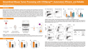 科学海报Streamlined Mouse Tumor Processing with STEMprep™: Automated, Efficient, and Reliable

 实验方案How to Process Leukocyte Reduction System (LRS) Cones/Chambers for Downstream Cell Isolation
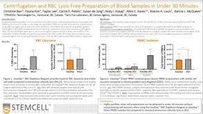 科学海报Centrifugation and RBC Lysis-Free Preparation of Blood Samples in Under 30 Minutes
 技术公告Simplify Your PBMC Isolations with the EasySep™ Direct Human PBMC Isolation Kit

 沪公网安备31010102008431号