Brandl M et al. (AUG 1999)
Experimental hematology 27 8 1264--70
Bispecific antibody fragments with CD20 X CD28 specificity allow effective autologous and allogeneic T-cell activation against malignant cells in peripheral blood and bone marrow cultures from patients with B-cell lineage leukemia and lymphoma.
Bispecific antibodies directed against tumor-associated target antigens and to surface receptors mediating T-cell activation,such as the TCR/CD3 complex and the costimulatory receptor CD28,are capable of mediating T-cell activation resulting in tumor cell killing. In this study,we used the B-cell-associated antigens CD19 and CD20 as target structures on human leukemic cells. We found that a combination of bispecific antibody fragments (bsFab2) with target x CD3 and target x CD28 specificity induces vigorous autologous T-cell activation and killing of malignant cells in peripheral blood and bone marrow cultures from patients with chronic lymphocytic leukemia and follicular lymphoma. The bsFab2 targeting CD20 were considerably more effective than those binding to CD19. The colony-forming capacity of treated bone marrow was impaired due to large amounts of tumor necrosis factor alpha produced during bsFab2-induced T-cell activation. Neutralizing tumor necrosis factor alpha antibodies were found to reverse this negative effect without affecting T-cell activation and tumor cell killing. CD20 x CD28 bsFab2,when used alone rather than in combination,markedly improved the recognition of leukemic cells by allogeneic T cells. Therefore,these reagents may be capable of enhancing the immunogenicity of leukemic cells in general and,in particular,of increasing the antileukemic activity of allogeneic donor buffy coat cells in relapsed bone marrow transplanted patients.
View Publication
产品号#:
04431
产品名:
MethoCult™ H4431
Yamashita J et al. (NOV 2000)
Nature 408 6808 92--6
Flk1-positive cells derived from embryonic stem cells serve as vascular progenitors.
Interaction between endothelial cells and mural cells (pericytes and vascular smooth muscle) is essential for vascular development and maintenance. Endothelial cells arise from Flk1-expressing (Flk1+) mesoderm cells,whereas mural cells are believed to derive from mesoderm,neural crest or epicardial cells and migrate to form the vessel wall. Difficulty in preparing pure populations of these lineages has hampered dissection of the mechanisms underlying vascular formation. Here we show that Flk1+ cells derived from embryonic stem cells can differentiate into both endothelial and mural cells and can reproduce the vascular organization process. Vascular endothelial growth factor promotes endothelial cell differentiation,whereas mural cells are induced by platelet-derived growth factor-BB. Vascular cells derived from Flk1+ cells can organize into vessel-like structures consisting of endothelial tubes supported by mural cells in three-dimensional culture. Injection of Flk1+ cells into chick embryos showed that they can incorporate as endothelial and mural cells and contribute to the developing vasculature in vivo. Our findings indicate that Flk1+ cells can act as 'vascular progenitor cells' to form mature vessels and thus offer potential for tissue engineering of the vascular system.
View Publication
产品号#:
06902
06952
00321
00322
00323
00324
00325
产品名:
Osada H et al. (APR 2001)
Transfusion 41 4 499--503
Detection of fetal HPCs in maternal circulation after delivery.
BACKGROUND: Circulation of mature fetal blood cells in the maternal blood for a certain postpartum period has been verified,but detailed study of the fetal HPCs has not been reported. The objective of this study was to evaluate the frequency and clearance of these cells in the peripheral blood of puerperal women. STUDY DESIGN AND METHODS: PBMNCs from 15 puerperal women who gave birth to male infants were cultured in semi-solid medium containing hematopoietic stimulating factors. Colonies formed in the medium were individually characterized,collected,and subjected to PCR amplification of the SRY gene on Y chromosome to confirm fetal origin. RESULTS: The mean numbers of fetal progenitor cell colonies isolated per mL of maternal blood were 1.63,2.48,0.56,0.12,and 0 on the day of delivery,at 4 days,1 month,6 months,and 1 year after delivery,respectively. There was no difference in the ratio of fetal versus maternal colonies between erythroid and granulocyte/macrophage lineages. CONCLUSION: The present study demonstrated that a significant number of fetal HPCs circulate in the maternal blood for a duration of at least 6 months after delivery.
View Publication
产品号#:
产品名:
Jasinski M et al. (OCT 2001)
Blood 98 7 2248--55
GATA1-Cre mediates Piga gene inactivation in the erythroid/megakaryocytic lineage and leads to circulating red cells with a partial deficiency in glycosyl phosphatidylinositol-linked proteins (paroxysmal nocturnal hemoglobinuria type II cells).
Patients with paroxysmal nocturnal hemoglobinuria (PNH) have blood cells deficient in glycosyl phosphatidylinositol (GPI)-linked proteins owing to a somatic mutation in the X-linked PIGA gene. To target Piga recombination to the erythroid/megakaryocytic lineage in mice,the Cre/loxP system was used,and Cre was expressed under the transcriptional regulatory sequences of GATA-1. Breeding of GATA1-cre (G) transgenic mice with mice carrying a floxed Piga (L) allele was associated with high embryonic lethality. However,double-transgenic (GL) mice that escaped early recombination looked healthy and were observed for 16 months. Flow cytometric analysis of peripheral blood cells showed that GL mice had up to 100% of red cells deficient in GPI-linked proteins. The loss of GPI-linked proteins on the cell surface occurred late in erythroid differentiation,causing a proportion of red cells to express low residual levels of GPI-linked proteins. Red cells with residual expression of GPI-linked proteins showed an intermediate sensitivity toward complement and thus resemble PNH type II cells in patients with PNH. Recombination of the floxed Piga allele was also detected in cultured megakaryocytes,mast cells,and eosinophils,but not in neutrophils,lymphocytes,or nonhematopoietic tissues. In summary,GATA1-Cre causes high-efficiency Piga gene inactivation in a GATA-1-specific pattern. For the first time,mice were generated that have almost 100% of red cells deficient in GPI-linked proteins. These animals will be valuable to further investigate the consequences of GPI-anchor deficiency on erythroid/megakaryocytic cells.
View Publication
产品号#:
05350
产品名:
Moreau-Gaudry F et al. (NOV 2001)
Blood 98 9 2664--72
High-level erythroid-specific gene expression in primary human and murine hematopoietic cells with self-inactivating lentiviral vectors.
Use of oncoretroviral vectors in gene therapy for hemoglobinopathies has been impeded by low titer vectors,genetic instability,and poor expression. Fifteen self- inactivating (SIN) lentiviral vectors using 4 erythroid promoters in combination with 4 erythroid enhancers with or without the woodchuck hepatitis virus postregulatory element (WPRE) were generated using the enhanced green fluorescent protein as a reporter gene. Vectors with high erythroid-specific expression in cell lines were tested in primary human CD34(+) cells and in vivo in the murine bone marrow (BM) transplantation model. Vectors containing the ankyrin-1 promoter showed high-level expression and stable proviral transmission. Two vectors containing the ankyrin-1 promoter and 2 erythroid enhancers (HS-40 plus GATA-1 or HS-40 plus 5-aminolevulinate synthase intron 8 [I8] enhancers) and WPRE expressed at levels higher than the HS2/beta-promoter vector in bulk unilineage erythroid cultures and individual erythroid blast-forming units derived from human BM CD34(+) cells. Sca1(+)/lineage(-) Ly5.1 mouse hematopoietic cells,transduced with these 2 ankyrin-1 promoter vectors,were injected into lethally irradiated Ly5.2 recipients. Eleven weeks after transplantation,high-level expression was seen from both vectors in blood (63%-89% of red blood cells) and erythroid cells in BM (70%-86% engraftment),compared with negligible expression in myeloid and lymphoid lineages in blood,BM,spleen,and thymus (0%-4%). The I8/HS-40-containing vector encoding a hybrid human beta/gamma-globin gene led to 43% to 113% human gamma-globin expression/copy of the mouse alpha-globin gene. Thus,modular use of erythroid-specific enhancers/promoters and WPRE in SIN-lentiviral vectors led to identification of high-titer,stably transmitted vectors with high-level erythroid-specific expression for gene therapy of red cell diseases.
View Publication

 EasySep™小鼠TIL(CD45)正选试剂盒


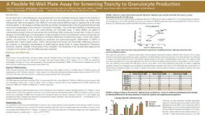 科学海报A Flexible 96-Well Plate Assay for Screening Toxicity to Granulocyte Production
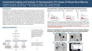 科学海报Automated Imaging and Analysis of Hematopoietic CFU Assays of Mouse Bone Marrow
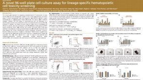 科学海报A Novel 96-well Plate Cell Culture Assay for Lineage-Specific Hematopoietic Cell Toxicity Screening
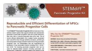
 科学海报STEMdiff™ Cerebral Organoid Kit: A New Tool for the Culture of 3D Brain Organoids Derived from hPSCs
 沪公网安备31010102008431号