Eirew P et al. (DEC 2008)
Nature medicine 14 12 1384--9
A method for quantifying normal human mammary epithelial stem cells with in vivo regenerative ability.
Previous studies have demonstrated that normal mouse mammary tissue contains a rare subset of mammary stem cells. We now describe a method for detecting an analogous subpopulation in normal human mammary tissue. Dissociated cells are suspended with fibroblasts in collagen gels,which are then implanted under the kidney capsule of hormone-treated immunodeficient mice. After 2-8 weeks,the gels contain bilayered mammary epithelial structures,including luminal and myoepithelial cells,their in vitro clonogenic progenitors and cells that produce similar structures in secondary transplants. The regenerated clonogenic progenitors provide an objective indicator of input mammary stem cell activity and allow the frequency and phenotype of these human mammary stem cells to be determined by limiting-dilution analysis. This new assay procedure sets the stage for investigations of mechanisms regulating normal human mammary stem cells (and possibly stem cells in other tissues) and their relationship to human cancer stem cell populations.
View Publication
产品号#:
05601
产品名:
EpiCult™-B 人培养基
Wang L-S et al. (FEB 2010)
Biomaterials 31 6 1148--57
Injectable biodegradable hydrogels with tunable mechanical properties for the stimulation of neurogenesic differentiation of human mesenchymal stem cells in 3D culture.
We report an injectable hydrogel scaffold system with tunable stiffness for controlling the proliferation rate and differentiation of human mesenchymal stem cells (hMSCs) in a three-dimensional (3D) context in normal growth media. The hydrogels composed of gelatin-hydroxyphenylpropionic acid (Gtn-HPA) conjugate were formed using the oxidative coupling of HPA moieties catalyzed by hydrogen peroxide (H(2)O(2)) and horseradish peroxidase (HRP). The stiffness of the hydrogels was readily tuned by varying the H(2)O(2) concentration without changing the concentration of polymer precursor. We found that the hydrogel stiffness strongly affected the cell proliferation rates. The rate of hMSC proliferation increased with the decrease in the stiffness of the hydrogel. Also,the neurogenesis of hMSCs was controlled by the hydrogel stiffness in a 3D context without the use of any additional biochemical signal. These cells which were cultured in hydrogels with lower stiffness for 3 weeks expressed much more neuronal protein markers compared to those cultured within stiffer hydrogels for the same period of time.
View Publication
产品号#:
05401
05402
05411
产品名:
MesenCult™ MSC 基础培养基(人)
MesenCult™ MSC 刺激补充剂(人)
MesenCult™ 增殖试剂盒(人)
Grajales L et al. (APR 2010)
Journal of molecular and cellular cardiology 48 4 735--45
Delayed enrichment of mesenchymal cells promotes cardiac lineage and calcium transient development.
Bone marrow-derived mesenchymal stem cells (BM-MSCs) can be induced to differentiate into myogenic cells. Despite their potential,previous studies have not been successful in producing a high percentage of cardiac-like cells with a muscle phenotype. We hypothesized that cardiac lineage development in BM-MSC is related to cell passage,culture milieu,and enrichment for specific cell subtypes before and during differentiation. Our study demonstrated that Lin(-) BM-MSC at an intermediate passage (IP; P8-P12) expressed cardiac troponin T (cTnT) after 21 days in culture. Cardiac TnT expression was similar whether IP cells were differentiated in media containing 5-azacytidine+2% FBS (AZA; 14%) or 2% FBS alone (LS; 12%) and both were significantly higher than AZA+5% FBS. This expression was potentiated by first enriching for CD117/Sca-1 cells followed by differentiation (AZA,39% and LS,28%). A second sequential enrichment for the dihydropyridine receptor subunit alpha2delta1 (DHPR-alpha2) resulted in cardiac TnT expressed in 54% of cultured cells compared to 28% of cells after CD117/Sca-1(+) enrichment. Cells enriched for CD117/Sca-1 and subjected to differentiation displayed spontaneous intracellular Ca(2+) transients with an increase in transient frequency and a 60% decrease in the transient duration amplitude between days 14 and 29. In conclusion,IP CD117/Sca-1(+) murine BM-MSCs display robust cardiac muscle lineage development that can be induced independent of AZA but is diminished under higher serum concentrations. Furthermore,temporal changes in calcium kinetics commensurate with increased cTnT expression suggest progressive maturation of a cardiac muscle lineage. Enrichment with CD117/Sca-1 to establish lineage commitment followed by DHPR-alpha2 in lineage developing cells may enhance the therapeutic potential of these cells for transplantation.
View Publication
产品号#:
19771
产品名:
EasySep™ 小鼠间充质干/祖细胞富集试剂盒
Dí et al. (DEC 2010)
Cardiovascular research 88 3 502--11
Endothelial progenitor cells undergo an endothelial-to-mesenchymal transition-like process mediated by TGFbetaRI.
AIMS: Endothelial progenitor cells (EPC) have been shown to repair pulmonary endothelium,although they can also migrate into the arterial intima and differentiate into smooth muscle-like (mesenchymal) cells contributing to intimal hyperplasia. The molecular mechanisms by which this process proceeds have not been fully elucidated. Here,we study whether genes involved in the endothelial-to-mesenchymal transition (EnMT) may contribute to the mesenchymal phenotype acquisition of EPC and we evaluate whether transforming growth factor β1 (TGFβ1) is involved in this process. METHODS AND RESULTS: Our results show that co-culture of EPC with smooth muscle cells (SMC) increases the expression of the mesenchymal cell markers α-smooth muscle actin,sm22-α,and myocardin,and decreases the expression of the endothelial cell marker CD31. In the same conditions,we also observed a concomitant increase in the gene expression of the EnMT-related transcription factors: slug,snail,zeb1,and endothelin-1. This indicates that mesenchymal phenotype acquisition occurred through an EnMT-like process. Inhibition of TGFβ receptor I (TGFβRI) downregulated snail gene expression,blocked the EnMT,and facilitated the differentiation of EPC to the endothelial cell lineage. Furthermore,TGFβRI inhibition decreased migration of EPC stimulated by SMC without affecting their functionality and adhesion capacity. CONCLUSION: These results indicate that EPC may differentiate into SMC-like cells through an EnMT-like process and that TGFβI plays an important role in the fate of EPC.
View Publication
Tauchmanovà et al. (MAY 2003)
Cancer 97 10 2453--61
Avascular necrosis in long-term survivors after allogeneic or autologous stem cell transplantation: a single center experience and a review.
BACKGROUND: The most debilitating skeletal complication of stem cell transplantation (SCT) is avascular necrosis (AVN). METHODS: Two hundred seven consecutive patients were evaluated prospectively for AVN. They survived disease free for more than 180 days after autologous or allogeneic SCT for hematologic malignancies. The diagnosis of AVN in suspicious cases was confirmed by magnetic resonance imaging. Possible correlations with treatments,bone mineral density (BMD),graft versus host disease (GVHD),and in vitro growth of fibroblast progenitors were investigated. Bone mineral density was evaluated by dual-energy X-ray absorptiometry in 100 transplanted patients,and the in vitro growth of fibroblast progenitors was monitored by a fibroblast colony-forming unit (CFU-F) assay in 30 patients after allogeneic SCT. RESULTS: Twelve patients developed AVN 3-114 months (median,26 months) following SCT: 10 (10%) after allogeneic SCT and 2 (1.9%) after autologous SCT (P = 0.04). Twenty-five joints were affected by AVN. All patients had femoral head involvement,which was managed with hip replacement in six of them. All but one patient who developed AVN after allogeneic SCT suffered from chronic GVHD (cGVHD). Avascular necrosis occurred 1-4 months after exacerbation or progression of cGVHD. Cumulative dose of steroids was similar in both SCT groups (including steroids given pretransplant for the basic disease),whereas treatment duration was significantly longer in the allogeneic SCT group. Avascular necrosis was related to the decreased number of bone marrow CFU-F colonies in vitro,but not to BMD values. CONCLUSIONS: Avascular necrosis is a skeletal complication that occurs more often after allogeneic than after autologous SCT. Occurrence of AVN symptoms after clinical follow-up of cGVHD suggests that cGVHD requiring long-term steroid therapy is one of the main risk factors for AVN. Avascular necrosis may be facilitated by a severe deficit in the repopulating capacity of bone marrow stromal stem cells after SCT.
View Publication
产品号#:
05401
05402
05411
产品名:
MesenCult™ MSC 基础培养基(人)
MesenCult™ MSC 刺激补充剂(人)
MesenCult™ 增殖试剂盒(人)
Rodrí et al. (MAY 2004)
Blood 103 9 3349--54
Interleukin-6 deficiency affects bone marrow stromal precursors, resulting in defective hematopoietic support.
Interleukin-6 (IL-6) is a critical factor in the regulation of stromal function and hematopoiesis. In vivo bromodeoxyuridine incorporation analysis indicates that the percentage of Lin(-)Sca-1(+) hematopoietic progenitors undergoing DNA synthesis is diminished in IL-6-deficient (IL-6(-/-)) bone marrow (BM) compared with wild-type BM. Reduced proliferation of IL-6(-/-) BM progenitors is also observed in IL-6(-/-) long-term BM cultures,which show defective hematopoietic support as measured by production of total cells,granulocyte macrophage-colony-forming units (CFU-GMs),and erythroid burst-forming units (BFU-Es). Seeding experiments of wild-type and IL-6(-/-) BM cells on irradiated wild-type or IL-6-deficient stroma indicate that the hematopoietic defect can be attributed to the stromal and not to the hematopoietic component. In IL-6(-/-) BM,stromal mesenchymal precursors,fibroblast CFUs (CFU-Fs),and stroma-initiating cells (SICs) are reduced to almost 50% of the wild-type BM value. Moreover,IL-6(-/-) stromata show increased CD34 and CD49e expression and reduced expression of the membrane antigens vascular cell adhesion molecule-1 (VCAM-1),Sca-1,CD49f,and Thy1. These data strongly suggest that IL-6 is an in vivo growth factor for mesenchymal precursors,which are in part implicated in the reduced longevity of the long-term repopulating stem cell compartment of IL-6(-/-) mice.
View Publication

 EasySep™小鼠TIL(CD45)正选试剂盒


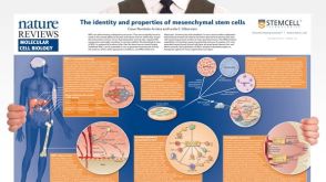 挂图The Identity and Properties of Mesenchymal Stem Cells Overview of MSC expansion, differentiation, immunoregulatory properties and therapeutic potential

 沪公网安备31010102008431号