Yamashita J et al. (NOV 2000)
Nature 408 6808 92--6
Flk1-positive cells derived from embryonic stem cells serve as vascular progenitors.
Interaction between endothelial cells and mural cells (pericytes and vascular smooth muscle) is essential for vascular development and maintenance. Endothelial cells arise from Flk1-expressing (Flk1+) mesoderm cells,whereas mural cells are believed to derive from mesoderm,neural crest or epicardial cells and migrate to form the vessel wall. Difficulty in preparing pure populations of these lineages has hampered dissection of the mechanisms underlying vascular formation. Here we show that Flk1+ cells derived from embryonic stem cells can differentiate into both endothelial and mural cells and can reproduce the vascular organization process. Vascular endothelial growth factor promotes endothelial cell differentiation,whereas mural cells are induced by platelet-derived growth factor-BB. Vascular cells derived from Flk1+ cells can organize into vessel-like structures consisting of endothelial tubes supported by mural cells in three-dimensional culture. Injection of Flk1+ cells into chick embryos showed that they can incorporate as endothelial and mural cells and contribute to the developing vasculature in vivo. Our findings indicate that Flk1+ cells can act as 'vascular progenitor cells' to form mature vessels and thus offer potential for tissue engineering of the vascular system.
View Publication
Bao F-XX et al. (APR 2016)
CNS neuroscience & therapeutics 22 8 648--660
Mitochondrial Membrane Potential-dependent Endoplasmic Reticulum Fragmentation is an Important Step in Neuritic Degeneration.
BACKGROUND Neuritic degeneration is an important early pathological step in neurodegeneration. AIM The purpose of this study was to explore the mechanisms connecting neuritic degeneration to the functional and morphological remodeling of endoplasmic reticulum (ER) and mitochondria. METHODS Here,we set up neuritic degeneration models by neurite cutting-induced neural degeneration in human-induced pluripotent stem cell-derived neurons. RESULTS We found that neuritic ER becomes fragmented and forms complexes with mitochondria,which induces IP3R-dependent mitochondrial Ca(2+) elevation and dysfunction during neuritic degeneration. Furthermore,mitochondrial membrane potential is required for ER fragmentation and mitochondrial Ca(2+) elevation during neuritic degeneration. Mechanically,tightening of the ER-mitochondria associations by expression of a short synthetic linker" and ER Ca(2+) releasing together could promote mitochondrial Ca(2+) elevation�
View Publication
产品号#:
05850
05857
05870
05875
85850
85857
85870
85875
产品名:
mTeSR™1
mTeSR™1
Yang Q et al. (NOV 2015)
Stem cell research 15 3 640--642
Human embryonic stem cells derived from abnormal blastocyst donated by Marfan syndrome patient.
Human embryonic stem cell (hESC) line was derived from abnormal blastocyst donated by Marfan syndrome patient after preimpantation genetic diagnosis (PGD) treatment. DNA sequencing analysis confirmed that the hESC line carried the heterozygous deletion mutation,c.3536delA,of FBN1 gene. Characteristic tests proved that the hESC line presented typicalmarkers of pluripotency and had the capability to formthe three germlayers both in vitro and in vivo.
View Publication
产品号#:
05850
05857
05870
05875
85850
85857
85870
85875
产品名:
mTeSR™1
mTeSR™1
Jang J et al. (APR 2016)
Cell 165 2 410--420
Primary Cilium-Autophagy-Nrf2 (PAN) Axis Activation Commits Human Embryonic Stem Cells to a Neuroectoderm Fate
Under defined differentiation conditions,human embryonic stem cells (hESCs) can be directed toward a mesendoderm (ME) or neuroectoderm (NE) fate,the first decision during hESC differentiation. Coupled with lineage-specific G1 lengthening,a divergent ciliation pattern emerged within the first 24 hr of induced lineage specification,and these changes heralded a neuroectoderm decision before any neural precursor markers were expressed. By day 2,increased ciliation in NE precursors induced autophagy that resulted in the inactivation of Nrf2 and thereby relieved transcriptional activation of OCT4 and NANOG. Nrf2 binds directly to upstream regions of these pluripotency genes to promote their expression and repress NE derivation. Nrf2 suppression was sufficient to rescue poorly neurogenic iPSC lines. Only after these events had been initiated did neural precursor markers get expressed at day 4. Thus,we have identified a primary cilium-autophagy-Nrf2 (PAN) control axis coupled to cell-cycle progression that directs hESCs toward NE.
View Publication
产品号#:
05850
05857
05870
05875
85850
85857
85870
85875
产品名:
mTeSR™1
mTeSR™1
Meng G et al. (APR 2016)
Methods in molecular biology (Clifton,N.J.)
An Effective and Reliable Xeno-free Cryopreservation Protocol for Single Human Pluripotent Stem Cells.
Efficient cryopreservation of human pluripotent stem cells (hPSCs) in chemically defined,xeno-free conditions is highly desirable for medical research and clinical applications such as cell-based therapies. Here we present a simple and effective slow freezing-rapid thawing protocol for the cryopreservation of feeder-free,single hPSCs. This cryopreservation protocol involves the supplementation of 10 % dimethyl sulfoxide (DMSO) and 10 $$M Rho-associated kinase inhibitor Y-27632 into two types of xeno-free,defined media supplements (Knockout Serum Replacement and TeSR2). High post-thaw cell recovery (˜90 %) and cell expansion (˜70 %) can be achieved using this protocol. The cryopreserved single cells retain the morphological characteristics of hPSCs and differentiation capabilities of pluripotent stem cells.
View Publication
产品号#:
05860
05880
产品名:
Agrawal P et al. (APR 2016)
ACS applied materials & interfaces 8 14 8870--8874
Fast, Efficient, and Gentle Transfection of Human Adherent Cells in Suspension
We demonstrate a highly efficient method for gene delivery into clinically relevant human cell types,such as induced pluripotent stem cells (iPSCs) and fibroblasts,reducing the protocol time by one full day. To preserve cell physiology during gene transfer,we designed a microfluidic strategy,which facilitates significant gene delivery in a short transfection time (textless1 min) for several human cell types. This fast,optimized and generally applicable cell transfection method can be used for rapid screening of different delivery systems and has significant potential for high-throughput cell therapy applications.
View Publication
Inhibition of class I histone deacetylases blunts cardiac hypertrophy through TSC2-dependent mTOR repression.
Altering chromatin structure through histone posttranslational modifications has emerged as a key driver of transcriptional responses in cells. Modulation of these transcriptional responses by pharmacological inhibition of class I histone deacetylases (HDACs),a group of chromatin remodeling enzymes,has been successful in blocking the growth of some cancer cell types. These inhibitors also attenuate the pathogenesis of pathological cardiac remodeling by blunting and even reversing pathological hypertrophy. The mechanistic target of rapamycin (mTOR) is a critical sensor and regulator of cell growth that,as part of mTOR complex 1 (mTORC1),drives changes in protein synthesis and metabolism in both pathological and physiological hypertrophy. We demonstrated through pharmacological and genetic methods that inhibition of class I HDACs suppressed pathological cardiac hypertrophy through inhibition of mTOR activity. Mice genetically silenced for HDAC1 and HDAC2 had a reduced hypertrophic response to thoracic aortic constriction (TAC) and showed reduced mTOR activity. We determined that the abundance of tuberous sclerosis complex 2 (TSC2),an mTOR inhibitor,was increased through a transcriptional mechanism in cardiomyocytes when class I HDACs were inhibited. In neonatal rat cardiomyocytes,loss of TSC2 abolished HDAC-dependent inhibition of mTOR activity,and increased expression of TSC2 was sufficient to reduce hypertrophy in response to phenylephrine. These findings point to mTOR and TSC2-dependent control of mTOR as critical components of the mechanism by which HDAC inhibitors blunt pathological cardiac growth. These results also suggest a strategy to modulate mTOR activity and facilitate the translational exploitation of HDAC inhibitors in heart disease.
View Publication
产品号#:
05850
05857
05870
05875
85850
85857
85870
85875
产品名:
mTeSR™1
mTeSR™1
Cui D et al. (APR 2016)
Bioscience,biotechnology,and biochemistry 80 8 1--8
Generating hESCs with reduced immunogenicity by disrupting TAP1 or TAPBP.
Human embryonic stem cells (hESCs) are thought to be a promising resource for cell therapy,while it has to face the major problem of graft immunological rejection. Major histocompatibility complex (MHC) class I expressed on the cell surface is the major cause of graft rejection. Transporter associated with antigen presentation 1 (TAP1) and TAP-associated glycoprotein (TAPBP) play important roles in regulating MHC class I expression. In this study,we generated TAP1- and TAPBP-deficient hESC lines,respectively,using transcription activator-like effector nucleases technique. These cells showed deficient expression of MHC class I on the cell surface and reduced immunogenicity compared with wild types,but maintained normal pluripotency,karyotypes,and differentiation ability. Thus,our findings are instrumental in developing a universal cell resource with both pluripotency and hypo-immunogenicity for transplantation therapy in the future.
View Publication
Lowe A et al. (MAY 2016)
Stem Cell Reports 6 5 743--756
Intercellular Adhesion-Dependent Cell Survival and ROCK-Regulated Actomyosin-Driven Forces Mediate Self-Formation of a Retinal Organoid
In this study we dissected retinal organoid morphogenesis in human embryonic stem cell (hESC)-derived cultures and established a convenient method for isolating large quantities of retinal organoids for modeling human retinal development and disease. Epithelialized cysts were generated via floating culture of clumps of Matrigel/hESCs. Upon spontaneous attachment and spreading of the cysts,patterned retinal monolayers with tight junctions formed. Dispase-mediated detachment of the monolayers and subsequent floating culture led to self-formation of retinal organoids comprising patterned neuroretina,ciliary margin,and retinal pigment epithelium. Intercellular adhesion-dependent cell survival and ROCK-regulated actomyosin-driven forces are required for the self-organization. Our data supports a hypothesis that newly specified neuroretina progenitors form characteristic structures in equilibrium through minimization of cell surface tension. In long-term culture,the retinal organoids autonomously generated stratified retinal tissues,including photoreceptors with ultrastructure of outer segments. Our system requires minimal manual manipulation,has been validated in two lines of human pluripotent stem cells,and provides insight into optic cup invagination in vivo.
View Publication

 EasySep™小鼠TIL(CD45)正选试剂盒


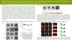 科学海报STEMdiff™ Cerebral Organoid Kit: A New Tool for the Culture of 3D Brain Organoids Derived from hPSCs

 沪公网安备31010102008431号