若您需要咨询产品或有任何技术问题,请通过官方电话 400 885 9050 或邮箱 info.cn@stemcell.com 与我们联系。

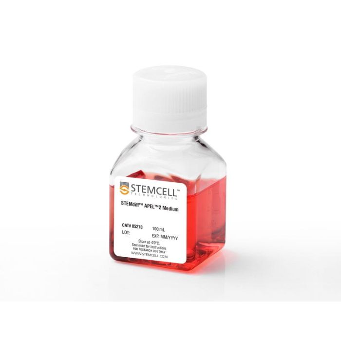
STEMdiff™ APEL™2 培养基

用于将人ES和iPS细胞分化为多种谱系的成分明确的无动物源培养基
只有 %1
¥2,466.00

产品号 #(选择产品)

产品号 #05270_C

用于将人ES和iPS细胞分化为多种谱系的成分明确的无动物源培养基

产品优势

  • 兼容 TeSR™ 培养的人类 ES 和 iPS 细胞
  • 兼容贴壁或胚体(EB)培养的分化方案
  • 在添加特定细胞因子或诱导因子时,能够支持内胚层、中胚层和外胚层的分化

总览

STEMdiff™ APEL™ 2 培养基是一种成分明确、无血清、无动物源的培养基,用于人胚胎干细胞 (ES) 和诱导多能干细胞 (iPS)的分化。该培养基基于 Andrew Elefanty 博士发表的 APEL 配方,不含任何诸如无蛋白杂交瘤培养基等不明确的成分。STEMdiff™ APEL™ 2 可用于贴壁或拟胚体 (EB) 的分化方案,例如与 AggreWell™ 配合使用。该培养基可以搭配多种不同的诱导因子或细胞因子使用,以支持向外胚层、中胚层和内胚层谱系的分化。

分类
专用培养基
 
细胞类型
多能干细胞
 
种属

 
应用
细胞培养,分化
 
品牌
STEMdiff
 
研究领域
干细胞生物学
 
制剂类别
无动物源
 

实验数据

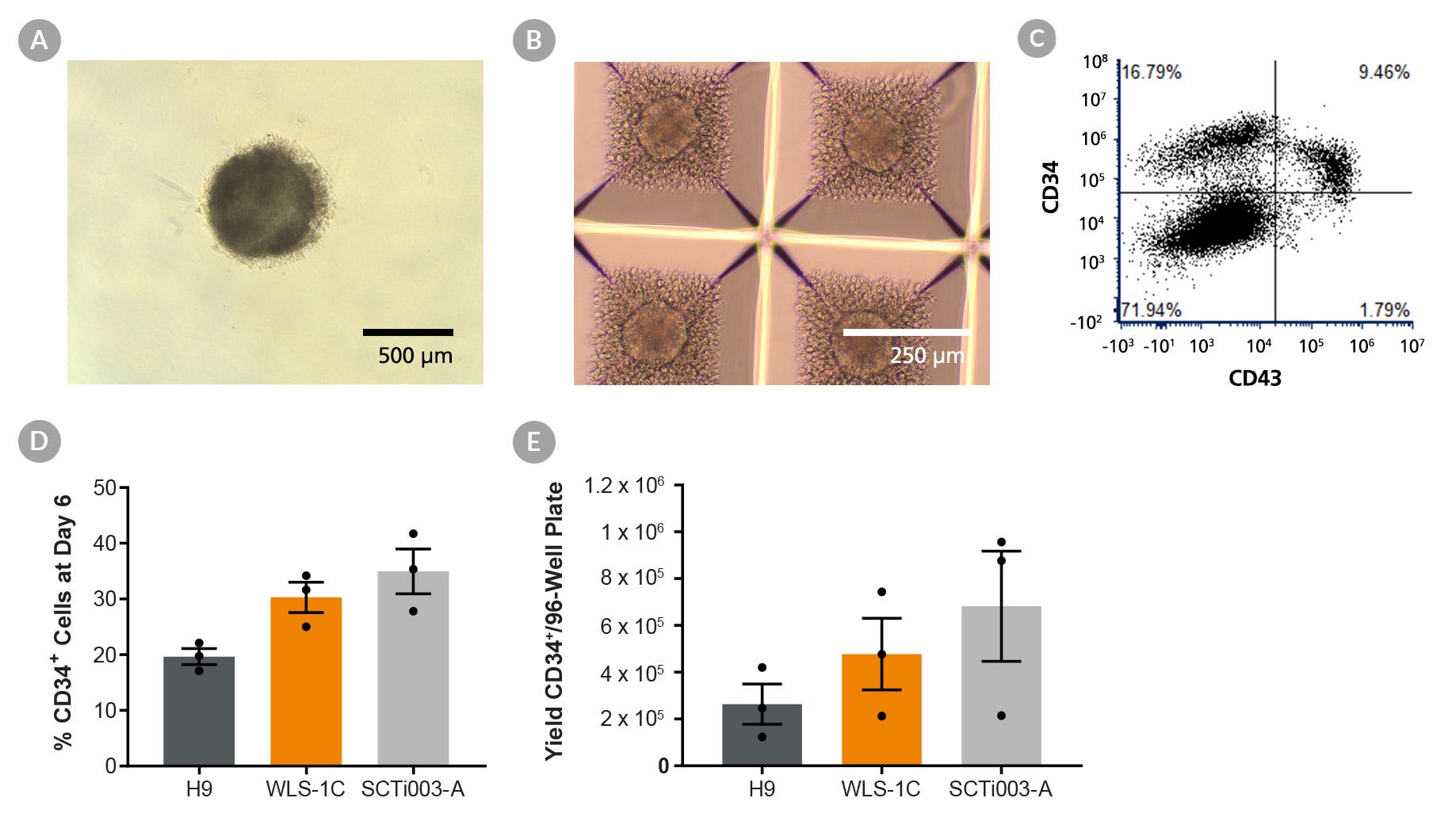
Figure 1. Generation of Hematopoietic Progenitors Using STEMdiff™ APEL™2 in a Spin EB Protocol

Figure 1. Generation of Hematopoietic Progenitors Using STEMdiff™ APEL™2 in a Spin EB Protocol

(A) Embryoid bodies can be generated by seeding 8000 hPSCs/well of a 96-well plate or (B) 500 hPSCs per microwell of AggreWell™400 24-well plates using STEMdiff™ APEL™2 Medium supplemented with Rho Kinase Inhibitor. Images shown were taken 24 hours after seeding. For hematopoietic differentiation using STEMdiff™ APEL™2 Medium, EBs in 96-well plates were generated following a modified Zhu and Kaufman protocol (STEMdiff™ APEL™2 supplemented with Human Recombinant SCF, ACF, Human Recombinant VEGF-165, ACF, Human Recombinant BMP-4, Rho Kinase Inhibitor IV, and Human Recombinant bFGF). At Day 6, EBs were dissociated, and cell counts and flow cytometry were performed. Hematopoietic progenitors were generated in one hESC (H9) and two hiPSC (SCTi003-A and WLS- 1C) lines. Cells were gated on singlets and viability. (C) Representative flow plot of WLS-1C. (D) Quantification of CD34+ cells at Day 6 and (E) quantification of yield of viable CD34+ cells generated per 96-well plate. Error bars are shown as mean +/- SEM, n = 3.

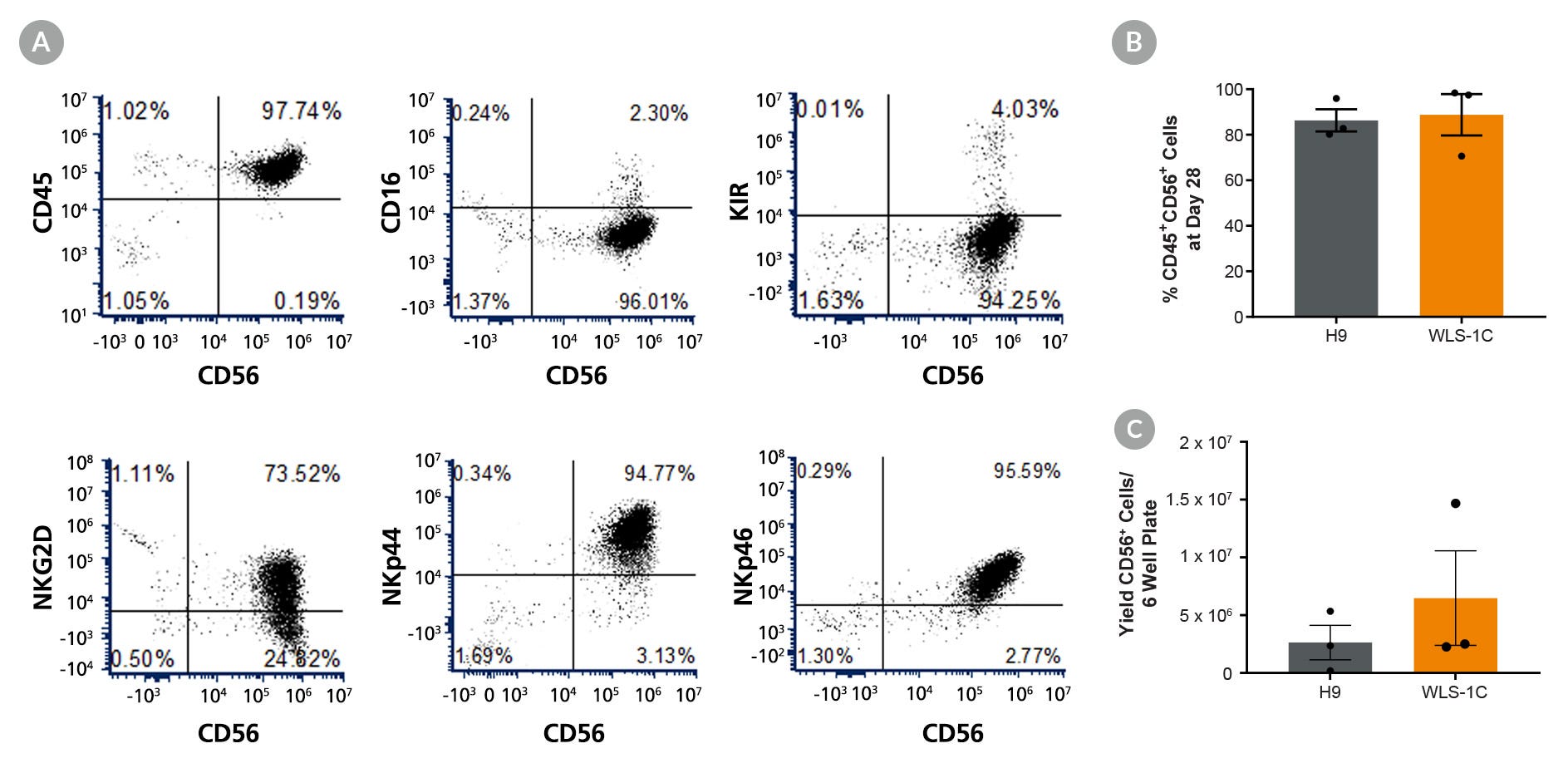
Figure 2. EBs Generated with STEMdiff™ APEL™2 Medium Can Differentiate to NK Cells at High Efficiency

Figure 2. EBs Generated with STEMdiff™ APEL™2 Medium Can Differentiate to NK Cells at High Efficiency

Following 6 days of hematopoietic differentiation in STEMdiff™ APEL™2 Medium (Figure 1), 16 EBs were transferred into each well of a gelatin-coated 6-well plate and cultured for 28 days in NK cell differentiation medium described in the Zhu and Kaufman protocol. After 28 days, floating cells were harvested, and cell counts and flow cytometry were performed using a panel of NK markers: Anti-Human CD56 (NCAM) Antibody, Clone HCD56 (APC), Anti-Human CD45 Antibody, Clone HI30 (PE), Anti-Human CD16 Antibody, Clone 3G8 (FITC), KIR (clone HP-MA4), NKG2D, NKp44, and NKp46. (A) Cell surface marker expression on pluripotent stem cell (PSC)-derived CD56+ NK Cells. Cells were gated on singlets and viability. NK cells were generated in 2 cell lines: ES cell line (H9) and human iPS cell line (WLS-1C). (B) Quantification of CD45+CD56+ cells at day 28 and (C) quantification of yield of viable CD56+ cells generated per 6-well plate. Error bars are shown as mean +/- SEM, n=3.

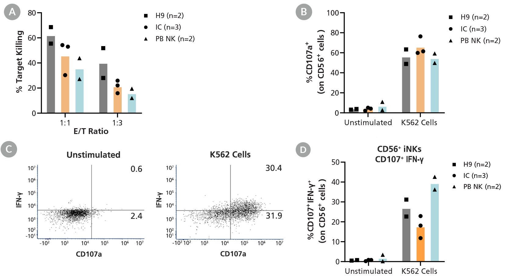
Figure 3. EBs Generated with STEMdiff™ APEL™2 Medium Can Differentiation Into Functional NK Cells

Figure 3. EBs Generated with STEMdiff™ APEL™2 Medium Can Differentiation Into Functional NK Cells

Following 6 days of hematopoietic differentiation in STEMdiff™ APEL™2 Medium (Figure 1), EBs were transferred to gelatin-coated plates and cultured for 28 days in NK differentiation media following the Kaufman et al. protocol. (A) After 28 days, hPSC-derived CD56+ NK cells were co-cultured with K562 target cells labeled with eBioscience™ Cell Proliferation Dye eFluor™ 670 for 5 hours at effector to target (E/T) ratios of 1:1 or 1:3. Positive controls were freshly isolated peripheral blood (PB) NK cells pre-cultured in ImmunoCult™ NK Cell Base Medium prior to co-culture with K562 target cells. K562 cells were cultured in the absence of NK cells as a negative control. The average percent target killing by hPSC-derived NK cells at a 1:1 E/T ratio ranged between 46% and 62%. For degranulation and IFN-γ production experiments, hPSC-derived CD56+ NK cells were co-cultured with K562 targets for 6 hours at an E/T ratio of 1:3, or left unstimulated in the absence of target cells. Co-cultures were set up in the presence of CD107a antibody, and monensin was added after 1 hour of co-culture. Cultures were stained with GloCell™ Fixable Viability Dye Red 780 and an anti-human CD56 antibody at the end of co-culture. (B) To measure degranulation, surface CD107a was assessed using flow cytometry. IFN-γ production was assessed following fixation and permeabilization of cells and staining with an antibody specific to human IFN-γ (clone 4S.B3). (C) Representative flow plots and (D) quantification show gating on fluorescently labeled CD56+CD107a+ NK cells. Upon stimulation, hPSC-derived CD56+ NK cells are able to degranulate, as shown by surface expression of CD107a (56 - 66% for K562 stimulation) and secrete IFN-γ (18 - 27% for K562 stimulation). Data are shown as mean among 2 - 3 independent experiments.

Figure 4. Generation of hPSC-Derived Hematopoietic Lineages Cultured in STEMdiff™ APEL™2 in 2D

Figure 4. Generation of hPSC-Derived Hematopoietic Lineages Cultured in STEMdiff™ APEL™2 in 2D

STEMdiff™ APEL™2 Medium can be used to generate multiple hematopoietic lineages from hPSCs in 2D when supplemented with appropriate cytokines. For example, Jeon et al. used STEMdiff™ APEL™2 Medium plus cytokines to generate CD34+/KDR+/CDH5+ hemogenic endothelial cells (not shown) and downstream erythroblasts. (A) By Day 18 of differentiation, red-colored erythroblasts were observed, and (B) by Day 30, flow cytometry showed a high proportion of cells were GlyA+/CD71- , markers of a mature erythrocyte population. Krisch et al. used STEMdiff™ APEL™2 Medium, StemSpan™-ACF Erythroid Expansion Medium, and Iscove's MDM plus cytokines to generate CD34+/CD31+ hemogenic endothelial cells (not shown) and downstream megakaryocyte cells. (C) Flow cytometry at Day 16 of differentiation demonstrated a CD41a+/CD42b+ cell population, markers of megakaryocyte cells. (D) By Day 21 of differentiation, immunostaining revealed expression of megakaryocyte markers and morphology typical of pro-platelets, indicated by the arrow. Adapted from Jeon et al. and Krisch et al., both available under a Creative Commons 4.0 License. In an alternative protocol, CD34+/CD45+ hematopoietic progenitors were generated by seeding hPSCs onto Matrigel® and culturing for 12 days in STEMdiff™ APEL™2 Medium supplemented with cytokines. On Day 12, hematopoietic progenitors were harvested from the culture supernatant, and cell counts and flow cytometry were performed. (E) Representative image of a hiPSCl line (WLS-1C) at Day 12 and (F) flow cytometry plot for hematopoietic progenitor markers CD34+CD45+. In this protocol, hematopoietic progenitors were generated from 3 cell lines; hESC line (H9) and hiPSC lines (SCTi003-A & WLS-1C). (G) Hematopoietic progenitor marker expression and (H) yield of live cells expressing CD34+CD45+ markers at Day 12 are shown.

Figure 5. Generation of hPSC-Derived Endothelial Cells Using STEMdiff™ APEL™2 Medium

Figure 5. Generation of hPSC-Derived Endothelial Cells Using STEMdiff™ APEL™2 Medium

hPSCs were plated at 50,000 cells/cm2 and cultured for 6 days in STEMdiff™ APEL™2 plus cytokines (BMP4, CHIR, VEGF) to generate endothelial cells based on the published Tan et al. 2D protocol. On Day 6, cells were harvested, and cell counts and flow cytometry were performed. Endothelial differentiation was performed in 3 cell lines, one hESC line (H9) and two hiPSC lines (SCTi003-A and WLS-1C ). (A) Representative image and (B) flow cytometry data for endothelial markers CD31+CD144+ of H9 at Day 6. Cells were gated on singlets and viability. (C) Quantification of CD31+CD144+ cells and (D) yield of viable CD31+CD144+ cells per cm2.

产品说明书及文档

请在《产品说明书》中查找相关支持信息和使用说明,或浏览下方更多实验方案。

Document Type
Product Name
Catalog #
Lot #
Language
Catalog #
05275, 05270
Lot #
All
Language
English
Document Type
Safety Data Sheet
Catalog #
05275, 05270
Lot #
All
Language
English

应用领域

本产品专为以下研究领域设计,适用于工作流程中的高亮阶段。探索这些工作流程,了解更多我们为各研究领域提供的其他配套产品。

相关材料与文献

技术资料 (5)

文献 (22)

Efficient derivation of lateral plate and paraxial mesoderm subtypes from human embryonic stem cells through GSKi-mediated differentiation. Tan JY et al. Stem cells and development 2013 JUL

Abstract

The vertebrae mesoderm is a source of cells that forms a variety of tissues,including the heart,vasculature,and blood. Consequently,the derivation of various mesoderm-specific cell types from human embryonic stem cells (hESCs) has attracted the interest of many investigators owing to their therapeutic potential in clinical applications. However,the need for efficient and reliable methods of differentiation into mesoderm lineage cell types remains a significant challenge. Here,we demonstrated that inhibition of glycogen synthase kinase-3 (GSK-3) is an essential first step toward efficient generation of the mesoderm. Under chemically defined conditions without additional growth factors/cytokines,short-term GSK inhibitor (GSKi) treatment effectively drives differentiation of hESCs into the primitive streak (PS),which can potentially commit toward the mesoderm when further supplemented with bone morphogenetic protein 4. Further analysis confirmed that the PS-like cells derived from GSKi treatment are bipotential,being able to specify toward the endoderm as well. Our findings suggest that the bipotential,PS/mesendoderm-like cell population exists only at the initial stages of GSK-3 inhibition,whereas long-term inhibition results in an endodermal fate. Lastly,we demonstrated that our differentiation approach could efficiently generate lateral plate (CD34(+)KDR(+)) and paraxial (CD34(-)PDGFRα(+)) mesoderm subsets that can be further differentiated along the endothelial and smooth muscle lineages,respectively. In conclusion,our study presents a unique approach for generating early mesoderm progenitors in a chemically directed fashion through the use of small-molecule GSK-3 inhibitor,which may be useful for future applications in regenerative medicine.
The epigenetic modifier ubiquitin-specific protease 22 (USP22) regulates embryonic stem cell differentiation via transcriptional repression of sex-determining region Y-box 2 (SOX2) Sussman RT et al. Journal of Biological Chemistry 2013 AUG

Abstract

Pluripotent embryonic stem cells (ESCs) undergo self-renewal until stimulated to differentiate along specific lineage pathways. Many of the transcriptional networks that drive reprogramming of a self-renewing ESC to a differentiating cell have been identified. However,fundamental questions remain unanswered about the epigenetic programs that control these changes in gene expression. Here we report that the histone ubiquitin hydrolase ubiquitin-specific protease 22 (USP22) is a critical epigenetic modifier that controls this transition from self-renewal to differentiation. USP22 is induced as ESCs differentiate and is necessary for differentiation into all three germ layers. We further report that USP22 is a transcriptional repressor of the locus encoding the core pluripotency factor sex-determining region Y-box 2 (SOX2) in ESCs,and this repression is required for efficient differentiation. USP22 occupies the Sox2 promoter and hydrolyzes monoubiquitin from ubiquitylated histone H2B and blocks transcription of the Sox2 locus. Our study reveals an epigenetic mechanism that represses the core pluripotency transcriptional network in ESCs,allowing ESCs to transition from a state of self-renewal into lineage-specific differentiation programs.
Erythropoietin critically regulates the terminal maturation of murine and human primitive erythroblasts Malik J et al. Haematologica 2013 NOV

Abstract

Primitive erythroid cells,the first red blood cells produced in the mammalian embryo,are necessary for embryonic survival. Erythropoietin and its receptor EpoR,are absolutely required for survival of late-stage definitive erythroid progenitors in the fetal liver and adult bone marrow. Epo- and Epor-null mice die at E13.5 with a lack of definitive erythrocytes. However,the persistence of circulating primitive erythroblasts raises questions about the role of erythropoietin/EpoR in primitive erythropoiesis. Using Epor-null mice and a novel primitive erythroid 2-step culture we found that erythropoietin is not necessary for specification of primitive erythroid progenitors. However,Epor-null embryos develop a progressive,profound anemia by E12.5 as primitive erythroblasts mature as a synchronous cohort. This anemia results from reduced primitive erythroblast proliferation associated with increased p27 expression,from advanced cellular maturation,and from markedly elevated rates of apoptosis associated with an imbalance in pro- and anti-apoptotic gene expression. Both mouse and human primitive erythroblasts cultured without erythropoietin also undergo accelerated maturation and apoptosis at later stages of maturation. We conclude that erythropoietin plays an evolutionarily conserved role in promoting the proliferation,survival,and appropriate timing of terminal maturation of primitive erythroid precursors.

更多信息

更多信息
物种
配方 无动物源性
质量保证:

产品仅供研究使用,不用于针对人或动物的诊断或治疗。 欲获悉更多关于STEMCELL的质控信息,请访问 STEMCELL.CN/COMPLIANCE.
Copyright © 2026 by STEMCELL Technologies. All rights reserved.

在线联系