
 EasySep™小鼠TIL(CD45)正选试剂盒


产品号 #17951_C
通过免疫磁珠负选分离无磁珠标记的人T细胞
若您需要咨询产品或有任何技术问题,请通过官方电话 400 885 9050 或邮箱 info.cn@stemcell.com 与我们联系。
通过免疫磁珠负选分离无磁珠标记的人T细胞
Isolating T cells doesn't have to take a long time. We developed this 8-minute T cell isolation kit so you can get to your downstream experiments sooner.

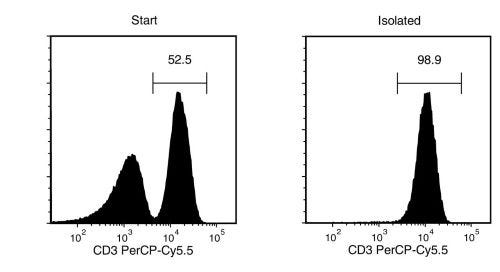
通过免疫磁珠负选从新鲜或冻存的人外周血单个核细胞 (PBMCs) 或裂解的白细胞单采术样本中分离出无磁珠标记和高纯度的T细胞。EasySep™无柱免疫磁珠分选技术结合单克隆抗体的特异性和无需分选柱的简便性,20多年来被广泛引用于已发表的文献中。
本EasySep™负选方案通过抗体复合物与磁珠标记非目标细胞,表达以下标志物的非目标细胞将被清除:CD14、CD16、CD19、CD36、CD56、CD66b、CD123及GlyA。通过EasySep™磁极将被磁珠标记的细胞与未被标记的目的细胞分离,接着只需将目的细胞倾倒或吸取至一个新的试管中,仅需8分钟即可获得高纯度的T细胞,且可立即用于流式细胞术、细胞培养或DNA/RNA提取等下游应用。
该产品可替代EasySep™人T细胞富集试剂盒 (产品号 #19051) 以进行更快的细胞分选。
如需从白细胞分离单采术中大规模分离人T细胞,请选用大规格(1x10^10细胞)试剂盒(产品号 #100-0695)
深入了解EasySep™免疫磁珠分选技术原理,或探索RoboSep™全自动免疫磁珠细胞分选方案。亦可选择即用型、符合伦理标准的冻存的人外周血pan-T细胞(使用EasySep™人T细胞分选试剂盒分离)。探索更多为您实验流程优化的产品,包括培养基、添加剂、抗体等。
磁极兼容性
• EasySep™磁极(产品号 #18000)
• “The Big Easy” EasySep™磁极(产品号 #18001)
• EasyPlate™ EasySep™磁极(产品号 #18102)
• Easy 50 EasySep™磁极(产品号 #18002)
• EasyEights™ EasySep™磁极(产品号 #18103)
• RoboSep™-S(产品号 #21000)
• Easy 250 EasySep™磁极(产品号 #100-0821)
分类
细胞分选试剂盒
细胞类型
T 细胞
种属
人
样本来源
白细胞单采术样本、PBMC
分选方法
负选
应用
细胞分选
品牌
EasySep,RoboSep
研究领域
嵌合体,HLA,免疫,细胞治疗开发
请在《产品说明书》中查找相关支持信息和使用说明,或浏览下方更多实验方案。
本产品专为以下研究领域设计,适用于工作流程中的高亮阶段。探索这些工作流程,了解更多我们为各研究领域提供的其他配套产品。
| Molecular Weight | 1645,1646 |
|---|---|
| 物种 | 人类 |
| Magnet Compatibility | • EasySep™ Magnet (Catalog #18000) • “The Big Easy” EasySep™ Magnet (Catalog #18001) • Easy 50 EasySep™ Magnet (Catalog #18002) • EasyPlate™ EasySep™ Magnet (Catalog 18102) • EasyEights™ EasySep™ Magnet (Catalog #18103) • RoboSep™-S (Catalog #21000) • Eas |
| 样本来源 | PBMC, 白细胞单采术样本 |
| Selection Method | Negative |
| Target | • EasySep™ Magnet (Catalog #18000) • “The Big Easy” EasySep™ Magnet (Catalog #18001) • Easy 50 EasySep™ Magnet (Catalog #18002) • EasyPlate™ EasySep™ Magnet (Catalog 18102) • EasyEights™ EasySep™ Magnet (Catalog #18103) • RoboSep™-S (Catalog #21000) • Eas |
| Pathway | Negative |

细胞解离试剂

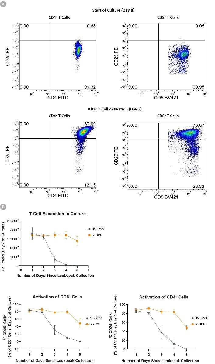
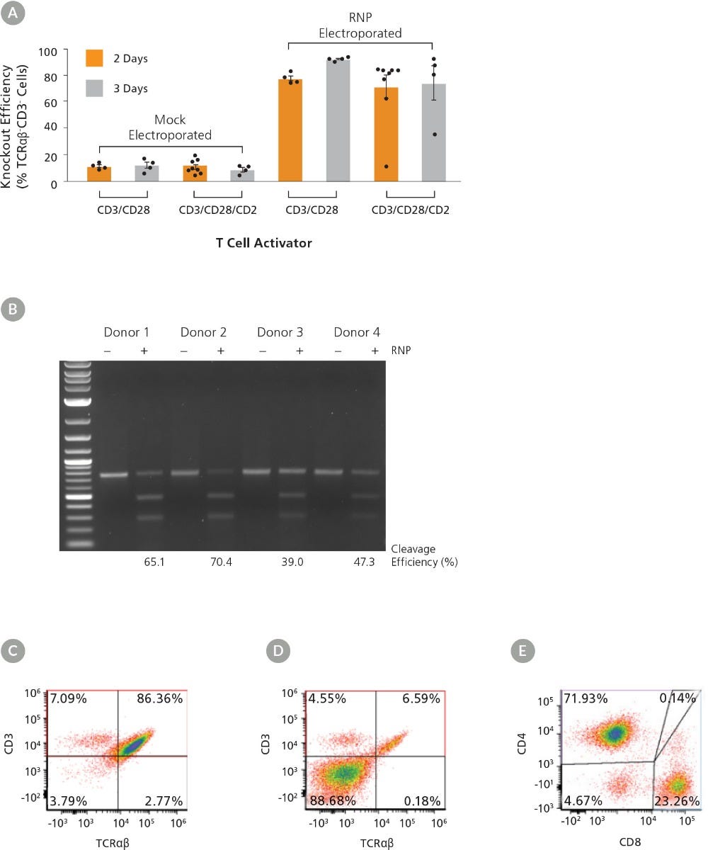
人T细胞激活扩增试剂

人T细胞激活扩增试剂
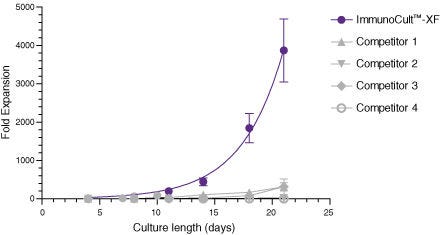
用于T细胞扩增的无血清和无异种成分培养基

小鼠Monoclonal IgG2b抗体,抗人、恒河猴、食蟹猴CD4

小鼠Monoclonal IgG1抗体,抗人、恒河猴、食蟹猴CD8a
小鼠(BALB/c)Monoclonal IgG1抗体,抗人、黑猩猩CD3
在线联系


 沪公网安备31010102008431号