若您需要咨询产品或有任何技术问题,请通过官方电话 400 885 9050 或邮箱 info.cn@stemcell.com 与我们联系。

MegaCult™-C胶原和含细胞因子培养基

胶原蛋白和含细胞因子的培养基,用于人CFU-Mk检测
只有 %1
¥16,676.00

产品号 #(选择产品)

产品号 #04961_C

胶原蛋白和含细胞因子的培养基,用于人CFU-Mk检测

产品优势

  • 无血清配方

产品组分包括

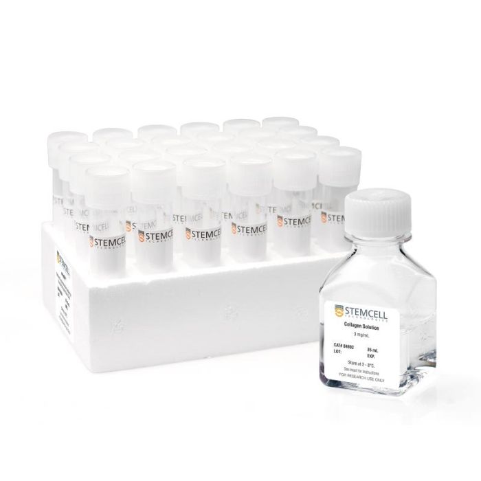
  • 胶原蛋白溶液,35 mL
  • MegaCult™-C含细胞因子培养基,24 × 2 mL(产品号 #04901)

总览

MegaCult™-C胶原和含细胞因子试剂盒包含进行人巨核细胞祖细胞(CFU-Mk)集落形成单位(CFU)检测所需的培养基和胶原蛋白溶液。

•MegaCult™-C含细胞因子培养基针对人骨髓、动员外周血和脐带血样本中的CFU-Mk生长进行优化,适用于CD34+富集的细胞、单个核细胞和采用其他纯化方法分离的细胞。培养基中含有重组人(rh) IL-3、IL-6和血小板生成素。

•胶原蛋白溶液用于制备胶原蛋白凝胶以及包被细胞培养表面。

以上组分也可以单独购买,以方便您的使用。

关于使用MegaCult™-C进行人CFU-Mk检测方法的更多信息,请参阅技术手册。

分类
半固体培养基,专用培养基
 
细胞类型
造血干/祖细胞
 
种属

 
应用
细胞培养,克隆筛选,功能学筛选
 
品牌
MegaCult
 
研究领域
药物发现与毒性检测,干细胞生物学
 
制剂类别
无血清
 

产品说明书及文档

请在《产品说明书》中查找相关支持信息和使用说明,或浏览下方更多实验方案。

Document Type
Product Name
Catalog #
Lot #
Language
Catalog #
04961
Lot #
All
Language
English
Catalog #
04961
Lot #
All
Language
English
Document Type
Technical Manual
Catalog #
04961
Lot #
All
Language
English
Document Type
Safety Data Sheet 1
Catalog #
04961
Lot #
All
Language
English
Document Type
Safety Data Sheet 2
Catalog #
04961
Lot #
All
Language
English

应用领域

本产品专为以下研究领域设计,适用于工作流程中的高亮阶段。探索这些工作流程,了解更多我们为各研究领域提供的其他配套产品。

相关材料与文献

技术资料 (2)

常见问题

Why is the MegaCult™-C formulation serum free?

MegaCult™-C is formulated without FBS to avoid inhibition of CFU-Mk growth by TGF beta and Platelet Factor-4, which are often present in the serum.

Why use semi-solid media?

Semi-solid media (such as methylcellulose-based or collagen-based) allow the clonal progeny of a single progenitor cell to stay together so you can recognize distinct colonies.

文献 (23)

Endogenous erythroid and megakaryocytic colony formation in serum-free, cytokine-free collagen gels. Dobo I et al. Journal of hematotherapy & stem cell research 1999 DEC

Abstract

We studied the suitability of collagen-based semisolid medium for assay of endogenous erythroid colony formation performed in myeloproliferative disorders. Bone marrow (BM) mononuclear cells (MNC) from 103 patients suspected of having polycythemia vera (PV,76 patients) or essential thrombocythemia (ET,27 patients) were grown in collagen-based,serum-free,cytokine-free semisolid medium. Colony analysis at day 8 or 10 showed that this collagen assay is specific,as endogenous growth of erythroid colonies was never observed in cultures of 16 healthy donors and 6 chronic myelogenous leukemia (CML) patients. Endogenous erythroid colony formation was observed in 53.3% of patients suspected of PV,with only 15.4% of positive cultures for patients with 1 minor PV criterion and 72% (p = 0.009) of positive cultures for patients with textgreater or =2 minor or 1 major PV criterion. Similarly,endogenous growth of erythroid colonies was found in 44.4% of patients suspected of ET,with 31.6% of positive cultures for patients with 1 ET criterion versus 75% for patients with textgreater or =2 ET criteria. In addition,we found that in collagen gels,tests of erythropoietin (EPO) hypersensitivity in the presence of 0.01 or 0.05 U/ml of EPO and tests of endogenous colony-forming units-megakaryocyte (CFU-MK) formation cannot be used to detect PV or ET,as these tests were positive for,respectively,21.4% and 50% of healthy donors and 83% and 50% of CML patients. A retrospective analysis suggests that collagen assays are more sensitive than methylcellulose assays to assess endogenous growth of erythroid colonies. In summary,serum-free collagen-based colony assays are simple and reliable assays of endogenous growth of erythroid colonies in myeloproliferative diseases. They also appear to be more sensitive than methylcellulose-based assays.
Comparison of four serum-free, cytokine-free media for analysis of endogenous erythroid colony growth in polycythemia vera and essential thrombocythemia. Dobo I et al. The hematology journal : the official journal of the European Haematology Association / EHA 2001 JAN

Abstract

INTRODUCTION: The assay of endogenous erythroid colony formation (EEC),a characteristic of polycythemia vera and essential thrombocythemia,is not standardized. In this multicentric study,we tested four semisolid,serum-free,cytokine-free media based on either methylcellulose (M1,M2) or collagen (C1,C2) commercialized for the EEC assay. MATERIALS AND METHODS: Bone marrow mononuclear cells (BMMC) from 73 individuals (62 patients with either polycythemia vera (26),essential thrombocythemia (19),secondary polyglobuly (17) or chronic myeloid leukemia (2) and 11 healthy donors) were grown in parallel in the four media without,or with 0.01 U/ml erythropoietin (EPo). RESULTS: In all four media EEC formation was specific,as it was not observed in cultures of patients with secondary polyglobuly or chronic myeloid leukemia,nor of healthy donors. Analysis of fresh or MGG-stained collagen gel cultures allowed detection of EEC formation significantly more frequently than methylcellulose-based media; addition of 0.01 U/ml of EPo had little or no effect on EEC formation. Collagen-based medium C1 gave better results than the other media tested: the 'C1' EEC assay was positive for 68.2% of polycythemia vera cultures with significantly higher median EEC numbers (6.5/10(5) BMMC for patients with one major criteria of polycythemia vera and 19 and 21/10(5) BMMC for patients with two or three major criteria,respectively). Medium C1 was also better for essential thrombocythemia cultures with 47.4% of positive results but with a low median EEC number (6.7/10(5) BMMC). When associated with the ELISA dosage of serum EPo,the 'C1' EEC assay allowed confirmation or elimination of the diagnosis of polycythemia vera for 91% (20/22) of polyglobulic patients. CONCLUSION: We propose that serum-free collagen-based culture systems be considered to standardize the EEC assay,now part of the new criteria of polycythemia vera.
Hematopoietic stem cell and progenitor defects in Sca-1/Ly-6A-null mice. Ito CY et al. Blood 2003 JAN

Abstract

Despite its wide use as a marker for hematopoietic stem cells (HSCs),the function of stem cell antigen-1 (Sca-1) (also known as lymphocyte activation protein-6A [Ly-6A]) in hematopoiesis remains poorly defined. We have previously established that Sca-1(-/-) T cells develop normally,although they are hyperresponsive to antigen. Here,we report detailed analysis of hematopoiesis in Sca-1-deficient animals. The differentiation potential of Sca-1-null bone marrow was determined from examination of the most mature precursors (culture colony-forming units [CFU-Cs]) to less committed progenitors (spleen CFUs [CFU-Ss]) to long-term repopulating HSCs. Sca-1-null mice are mildly thrombocytopenic with a concomitant decrease in megakaryocytes and their precursors. Bone marrow cells derived from Sca-1(-/-) mice also have decreased multipotential granulocyte,erythroid,macrophage,and megakaryocyte CFU (GEMM-CFU) and CFU-S progenitor activity. Competitive repopulation assays demonstrated that Sca-1(-/-) HSCs are at a competitive disadvantage compared with wild-type HSCs. To further analyze the potential of Sca-1(-/-) HSCs,serial transplantations were performed. While secondary repopulations using wild-type bone marrow completely repopulated Sca-1(-/-) mice,Sca-1(-/-) bone marrow failed to rescue one third of lethally irradiated wild-type mice receiving secondary bone marrow transplants from irradiation-induced anemia and contributed poorly to the surviving transplant recipients. These data strongly suggest that Sca-1 is required for regulating HSC self-renewal and the development of committed progenitor cells,megakaryocytes,and platelets. Thus,our studies conclusively demonstrate that Sca-1,in addition to being a marker of HSCs,regulates the developmental program of HSCs and specific progenitor populations.

更多信息

更多信息
物种
配方 无血清
质量保证:

产品仅供研究使用,不用于针对人或动物的诊断或治疗。
Copyright © 2026 by STEMCELL Technologies. All rights reserved.

在线联系