Lin H et al. (MAR 2009)
Experimental biology and medicine (Maywood,N.J.) 234 3 342--53
Maitake beta-glucan enhances umbilical cord blood stem cell transplantation in the NOD/SCID mouse.
Beta glucans are cell wall constituents of yeast,fungi and bacteria,as well as mushrooms and barley. Glucans are not expressed on mammalian cells and are recognized as pathogen-associated molecular patterns (PAMPS) by pattern recognition receptors (PRR). Beta glucans have potential activity as biological response modifiers for hematopoiesis and enhancement of bone marrow recovery after injury. We have reported that Maitake beta glucan (MBG) enhanced mouse bone marrow (BMC) and human umbilical cord blood (CB) cell granulocyte-monocyte colony forming unit (GM-CFU) activity in vitro and protected GM-CFU forming stem cells from doxorubicin (DOX) toxicity. The objective of this study was to determine the effects of MBG on expansion of phenotypically distinct subpopulations of progenitor and stem cells in CB from full-term infants cultured ex vivo and on homing and engraftment in vivo in the nonobese diabetic/severe combined immunodeficient (NOD/SCID) mouse. MBG promoted a greater expansion of CD34+CD33+CD38- human committed hematopoietic progenitor (HPC) cells compared to the conventional stem cell culture medium (P = 0.002 by ANOVA). CD34+CXCR4+CD38- early,uncommitted human hematopoietic stem cell (HSC) numbers showed a trend towards increase in response to MBG. The fate of CD34+ enriched CB cells after injection into the sublethally irradiated NOS/SCID mouse was evaluated after retrieval of xenografted human CB from marrow and spleen by flow cytometric analysis. Oral administration of MBG to recipient NOS/SCID mice led to enhanced homing at 3 days and engraftment at 6 days in mouse bone marrow (P = 0.002 and P = 0.0005,respectively) compared to control mice. More CD34+ human CB cells were also retrieved from mouse spleen in MBG treated mice at 6 days after transplantation. The studies suggest that MBG promotes hematopoiesis through effects on CD34+ progenitor cell expansion ex vivo and when given to the transplant recipient could enhance CD34+ precursor cell homing and support engraftment.
View Publication
产品类型:
产品号#:
02690
09600
09650
09850
15026
15066
产品名:
StemSpan™ CC100
StemSpan™ SFEM
StemSpan™ SFEM
RosetteSep™人造血祖细胞富集抗体混合物
RosetteSep™人造血祖细胞富集抗体混合物
Avitabile D et al. (MAY 2011)
American journal of physiology. Heart and circulatory physiology 300 5 H1875--84
Human cord blood CD34+ progenitor cells acquire functional cardiac properties through a cell fusion process.
The efficacy of cardiac repair by stem cell administration relies on a successful functional integration of injected cells into the host myocardium. Safety concerns have been raised about the possibility that stem cells may induce foci of arrhythmia in the ischemic myocardium. In a previous work (36),we showed that human cord blood CD34(+) cells,when cocultured on neonatal mouse cardiomyocytes,exhibit excitation-contraction coupling features similar to those of cardiomyocytes,even though no human genes were upregulated. The aims of the present work are to investigate whether human CD34(+) cells,isolated after 1 wk of coculture with neonatal ventricular myocytes,possess molecular and functional properties of cardiomyocytes and to discriminate,using a reporter gene system,whether cardiac differentiation derives from a (trans)differentiation or a cell fusion process. Umbilical cord blood CD34(+) cells were isolated by a magnetic cell sorting method,transduced with a lentiviral vector carrying the enhanced green fluorescent protein (EGFP) gene,and seeded onto primary cultures of spontaneously beating rat neonatal cardiomyocytes. Cocultured EGFP(+)/CD34(+)-derived cells were analyzed for their electrophysiological features at different time points. After 1 wk in coculture,EGFP(+) cells,in contact with cardiomyocytes,were spontaneously contracting and had a maximum diastolic potential (MDP) of -53.1 mV,while those that remained isolated from the surrounding myocytes did not contract and had a depolarized resting potential of -11.4 mV. Cells were then resuspended and cultured at low density to identify EGFP(+) progenitor cell derivatives. Under these conditions,we observed single EGFP(+) beating cells that had acquired an hyperpolarization-activated current typical of neonatal cardiomyocytes (EGFP(+) cells,-2.24 ± 0.89 pA/pF; myocytes,-1.99 ± 0.63 pA/pF,at -125 mV). To discriminate between cell autonomous differentiation and fusion,EGFP(+)/CD34(+) cells were cocultured with cardiac myocytes infected with a red fluorescence protein-lentiviral vector; under these conditions we found that 100% of EGFP(+) cells were also red fluorescent protein positive,suggesting cell fusion as the mechanism by which cardiac functional features are acquired.
View Publication
产品类型:
产品号#:
09600
09650
产品名:
StemSpan™ SFEM
StemSpan™ SFEM
Liu C et al. (DEC 2010)
Blood 116 25 5518--27
Progenitor cell dose determines the pace and completeness of engraftment in a xenograft model for cord blood transplantation.
Two critical concerns in clinical cord blood transplantation are the initial time to engraftment and the subsequent restoration of immune function. These studies measured the impact of progenitor cell dose on both the pace and strength of hematopoietic reconstitution by transplanting nonobese diabetic/severe combined immunodeficiency/interleukin-2 receptor-gamma-null (NSγ) mice with lineage-depleted aldehyde dehydrogenase-bright CD34(+) human cord blood progenitors. The progress of each transplant was monitored over an extended time course by repeatedly analyzing the peripheral blood for human hematopoietic cells. In vivo human hematopoietic development was complete. After long-term transplantation assays (≥ 19 weeks),human T-cell development was documented within multiple tissues in 16 of 32 NSγ mice. Human T-cell differentiation was active within NSγ thymuses,as documented by the presence of CD4(+) CD8(+) T-cell progenitors as well as T-cell receptor excision circles. It is important to note that although myeloid and B-cell engraftment was detected as early as 4 weeks after transplantation,human T-cell development was exclusively late onset. High progenitor cell doses were associated with a robust human hematopoietic chimerism that accelerated both initial time to engraftment and subsequent T-cell development. At lower progenitor cell doses,the chimerism was weak and the human hematopoietic lineage development was frequently incomplete.
View Publication
产品类型:
产品号#:
01700
01705
01701
01702
14056
14066
28600
19056
19056RF
19756
19756RF
产品名:
ALDEFLUOR™ 试剂盒
ALDEFLUOR™ DEAB试剂, 1.5 mM, 1 mL
ALDEFLUOR™检测缓冲液
L-Calc™有限稀释软件
Orlandi A et al. (APR 2008)
American journal of physiology. Heart and circulatory physiology 294 4 H1541--9
Functional properties of cells obtained from human cord blood CD34+ stem cells and mouse cardiac myocytes in coculture.
Prior in vitro studies suggested that different types of hematopoietic stem cells may differentiate into cardiomyocytes. The present work examined whether human CD34(+) cells from the human umbilical cord blood (hUCB),cocultured with neonatal mouse cardiomyocytes,acquire the functional properties of myocardial cells and express human cardiac genes. hUCB CD34(+) cells were cocultured onto cardiomyocytes following an infection with a lentivirus-encoding enhanced green fluorescent protein (EGFP). After 7 days,mononucleated EGFP(+) cells were tested for their electrophysiological features by patch clamp and for cytosolic [Ca(2+)] ([Ca(2+)](i)) homeostasis by [Ca(2+)](i) imaging of X-rhod1-loaded cells. Human Nkx2.5 and GATA-4 expression was examined in cocultured cell populations by real-time RT-PCR. EGFP(+) cells were connected to surrounding cells by gap junctions,acquired electrophysiological properties similar to those of cardiomyocytes,and showed action potential-associated [Ca(2+)](i) transients. These cells also exhibited spontaneous sarcoplasmic reticulum [Ca(2+)](i) oscillations and the associated membrane potential depolarization. However,RT-PCR of both cell populations showed no upregulation of human-specific cardiac genes. In conclusion,under our experimental conditions,hUCB CD34(+) cells cocultured with murine cardiomyocytes formed cells that exhibited excitation-contraction coupling features similar to those of cardiomyocytes. However,the expression of human-specific cardiac genes was undetectable by RT-PCR.
View Publication


 EasySep™小鼠TIL(CD45)正选试剂盒




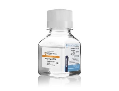 CryoStor®CSB 无动物成分和无蛋白质冷冻保存配方
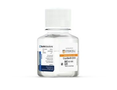 CryoStor® CS10 以cGMP标准生产、不含动物成分、成分明确、含10% DMSO的冻存液
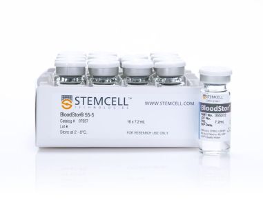 BloodStor®55-5 优化的血源细胞和组织生物保存试剂
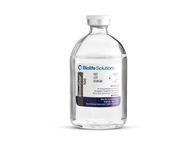 BloodStor®100 细胞和组织的生物保存试剂
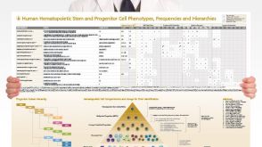 挂图Human Hematopoietic Stem and Progenitor Cell Phenotyping Overview of subset surface markers, frequencies and assays for analysis


 沪公网安备31010102008431号