B. L. Jamison et al. (jul 2019)
Journal of immunology (Baltimore,Md. : 1950) 203 1 48--57
Nanoparticles Containing an Insulin-ChgA Hybrid Peptide Protect from Transfer of Autoimmune Diabetes by Shifting the Balance between Effector T Cells and Regulatory T Cells.
CD4 T cells play a critical role in promoting the development of autoimmunity in type 1 diabetes. The diabetogenic CD4 T cell clone BDC-2.5,originally isolated from a NOD mouse,has been widely used to study the contribution of autoreactive CD4 T cells and relevant Ags to autoimmune diabetes. Recent work from our laboratory has shown that the Ag for BDC-2.5 T cells is a hybrid insulin peptide (2.5HIP) consisting of an insulin C-peptide fragment fused to a peptide from chromogranin A (ChgA) and that endogenous 2.5HIP-reactive T cells are major contributors to autoimmune pathology in NOD mice. The objective of this study was to determine if poly(lactide-co-glycolide) (PLG) nanoparticles (NPs) loaded with the 2.5HIP Ag (2.5HIP-coupled PLG NPs) can tolerize BDC-2.5 T cells. Infusion of 2.5HIP-coupled PLG NPs was found to prevent diabetes in an adoptive transfer model by impairing the ability of BDC-2.5 T cells to produce proinflammatory cytokines through induction of anergy,leading to an increase in the ratio of Foxp3+ regulatory T cells to IFN-gamma+ effector T cells. To our knowledge,this work is the first to use a hybrid insulin peptide,or any neoepitope,to re-educate diabetogenic T cells and may have significant implications for the development of an Ag-specific therapy for type 1 diabetes patients.
View Publication
产品类型:
产品号#:
19852
19852RF
18783
18783RF
18765
18765RF
产品名:
EasySep™小鼠CD4+ T细胞分选试剂盒
RoboSep™ 小鼠CD4+ T细胞分选试剂盒
EasySep™小鼠CD4+CD25+调节性T细胞分选试剂盒II
RoboSep™ 小鼠CD4+CD25+调节性T细胞分选试剂盒II
EasySep™小鼠CD4+ CD62L+ T细胞分选试剂盒
RoboSep™ 小鼠CD4+ CD62L+ T细胞分选试剂盒
Allan LL et al. (SEP 2009)
Blood 114 12 2411--6
Apolipoprotein-mediated lipid antigen presentation in B cells provides a pathway for innate help by NKT cells.
Natural killer T (NKT) cells are innate-like lymphocytes that recognize lipid antigens and have been shown to enhance B-cell activation and antibody production. B cells typically recruit T-cell help by presenting internalized antigens recognized by their surface antigen receptor. Here,we demonstrate a highly efficient means whereby human B cells present lipid antigens to NKT cells,capturing the antigen using apolipoprotein E (apoE) and the low-density lipoprotein receptor (LDL-R). ApoE dramatically enhances B-cell presentation of alpha-galactosylceramide (alphaGalCer),an exogenous CD1d presented antigen,inducing activation of NKT cells and the subsequent activation of B cells. B cells express the LDL-R on activation,and the activation of NKT cells by B cells is completely LDL-R dependent,as shown by blocking experiments and the complete lack of presentation when using apoE2,an isoform of apoE incapable of LDL-R binding. The dependence on apoE and the LDL-R is much more pronounced in B cells than we had previously seen in dendritic cells,which can apparently use alternate pathways of lipid antigen uptake. Thus,B cells use an apolipoprotein-mediated pathway of lipid antigen presentation,which constitutes a form of innate help for B cells by NKT cells.
View Publication
Christopher MJ et al. (FEB 2011)
The Journal of experimental medicine 208 2 251--60
Expression of the G-CSF receptor in monocytic cells is sufficient to mediate hematopoietic progenitor mobilization by G-CSF in mice.
Granulocyte colony-stimulating factor (G-CSF),the prototypical mobilizing cytokine,induces hematopoietic stem and progenitor cell (HSPC) mobilization from the bone marrow in a cell-nonautonomous fashion. This process is mediated,in part,through suppression of osteoblasts and disruption of CXCR4/CXCL12 signaling. The cellular targets of G-CSF that initiate the mobilization cascade have not been identified. We use mixed G-CSF receptor (G-CSFR)-deficient bone marrow chimeras to show that G-CSF-induced mobilization of HSPCs correlates poorly with the number of wild-type neutrophils. We generated transgenic mice in which expression of the G-CSFR is restricted to cells of the monocytic lineage. G-CSF-induced HSPC mobilization,osteoblast suppression,and inhibition of CXCL12 expression in the bone marrow of these transgenic mice are intact,demonstrating that G-CSFR signals in monocytic cells are sufficient to induce HSPC mobilization. Moreover,G-CSF treatment of wild-type mice is associated with marked loss of monocytic cells in the bone marrow. Finally,we show that bone marrow macrophages produce factors that support the growth and/or survival of osteoblasts in vitro. Together,these data suggest a model in which G-CSFR signals in bone marrow monocytic cells inhibit the production of trophic factors required for osteoblast lineage cell maintenance,ultimately leading to HSPC mobilization.
View Publication
Wognum AW et al. ( )
Archives of medical research 34 6 461--75
Identification and isolation of hematopoietic stem cells.
Hematopoietic stem cells (HSCs) are defined by their ability to repopulate all of the hematopoietic lineages in vivo and sustain the production of these cells for the life span of the individual. In the absence of reliable direct markers for HSCs,their identification and enumeration depends on functional long-term,multilineage,in vivo repopulation assays. The extremely low frequency of HSCs in any tissue and the absence of a specific HSC phenotype have made their purification and characterization a highly challenging goal. HSCs and primitive hematopoietic cells can be distinguished from mature blood cells by their lack of lineage-specific markers and presence of certain other cell-surface antigens,such as CD133 (for human cells) and c-kit and Sca-1 (for murine cells). Functional analyses of purified subpopulations of primitive hematopoietic cells have led to the development of several procedures for isolating cell populations that are highly enriched in cells with in vivo stem cell activity. Simplified methods for obtaining these cells at high yield have been important to the practical exploitation of such advances. This article reviews recent progress in identifying human and mouse HSCs and current techniques for their purification.
View Publication
Yeo C et al. (SEP 2009)
Regenerative Medicine 4 5 689--696
Ficoll-Paque™ versus Lymphoprep™: a comparative study of two density gradient media for therapeutic bone marrow mononuclear cell preparations
AIMS Contradictory outcomes from recent clinical trials investigating the transplantation of autologous bone marrow mononuclear cell (BM-MNC) fraction containing stem/progenitor cells to damaged myocardium,following acute myocardial infarction,may be,in part,due to the different cell isolation protocols used. We compared total BM-MNC numbers and its cellular subsets obtained following isolation using Ficoll-Paque and Lymphoprep - two different density gradient media used in the clinical trials. MATERIALS & METHODS Bone marrow samples were taken from patients entered into the REGENERATE-IHD clinical trial after 5 days of subcutaneous granulocyte colony-stimulating factor injections. Each sample was divided equally for BM-MNC isolation using Ficoll-Paque and Lymphoprep,keeping all other procedural steps constant. Isolated fractions were characterized for hematopoietic stem cells,endothelial progenitor cells,T lymphocytes,B lymphocytes and NK cells using cell surface markers CD34(+),CD133(+)VEGFR2(+),CD45(+)CD3(+),CD45(+)CD19(+) and CD45(+)CD16(+)CD56(+),respectively. There were no significant differences in the absolute numbers and percentage cell recovery of various mononuclear cell types recovered following separation using either density gradient media. Cell viability and the proportion of various cell phenotypes investigated were similar between the two media. They were also equally efficient in excluding unwanted red blood cells,granulocytes and platelets from the final cell products. CONCLUSION We demonstrated that the composition and quantity of cell types found within therapeutic BM-MNC preparations for use in clinical trials of cardiac stem cell transplantation are not influenced by the type of density gradient media used when comparing Ficoll-Paque and Lymphoprep.
View Publication

 EasySep™小鼠TIL(CD45)正选试剂盒


 技术公告Isolate Mouse CD45.1 or CD45.2 Positive Cells with EasySep™ Release Mouse Positive Selection Kits
 27:26
线上讲座How to Optimize Your T Cell Therapy Workflow—Without the Use of Serum or Feeder Cells发布日期: 08/27/2024
27:26
线上讲座How to Optimize Your T Cell Therapy Workflow—Without the Use of Serum or Feeder Cells发布日期: 08/27/2024 科学海报Optimized CRISPR-Cas9 Editing of Primary Human Immune and CD34+ Hematopoietic Stem and Progenitor Cells using a Mechanoporation Platform
 技术公告Generation of Natural Killer Cells from Human Pluripotent Stem Cells Using STEMdiff™ and StemSpan™ Media and Supplements
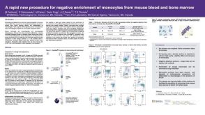 科学海报Procedure for Negative Enrichment of Monocytes from Mouse Blood and Bone Marrow
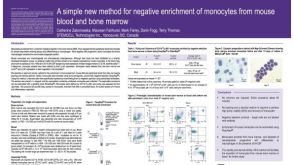 科学海报Method for Negative Enrichment of Monocytes from Mouse Blood and Bone Marrow
 沪公网安备31010102008431号