
 EasySep™小鼠TIL(CD45)正选试剂盒


产品号 #05320_C
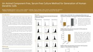
用于将人多能干细胞(hPSCs)分化为单核细胞
若您需要咨询产品或有任何技术问题,请通过官方电话 400 885 9050 或邮箱 info.cn@stemcell.com 与我们联系。
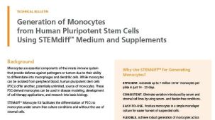
用于将人多能干细胞(hPSCs)分化为单核细胞
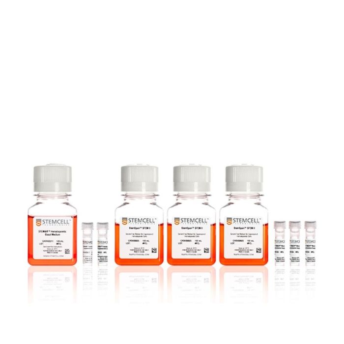
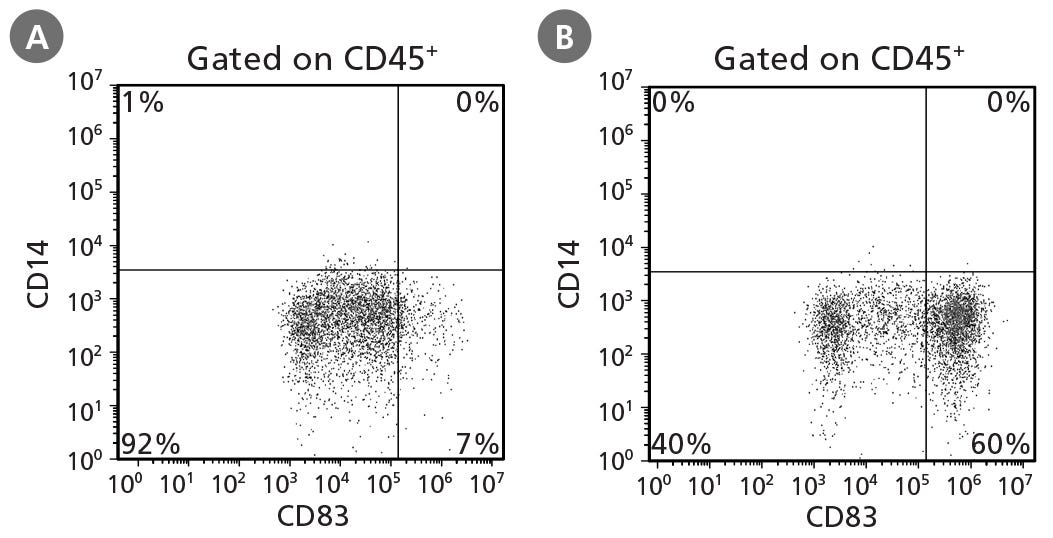
使用无饲养层、无血清的 STEMdiff™ 单核细胞试剂盒,可将人多能干细胞(hPSCs)分化为表达 CD14 的单核细胞。
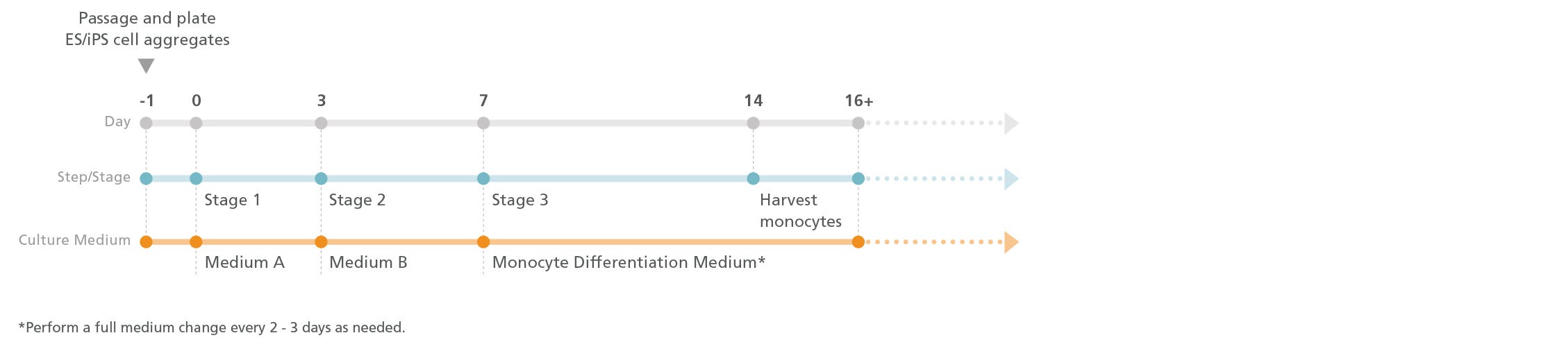
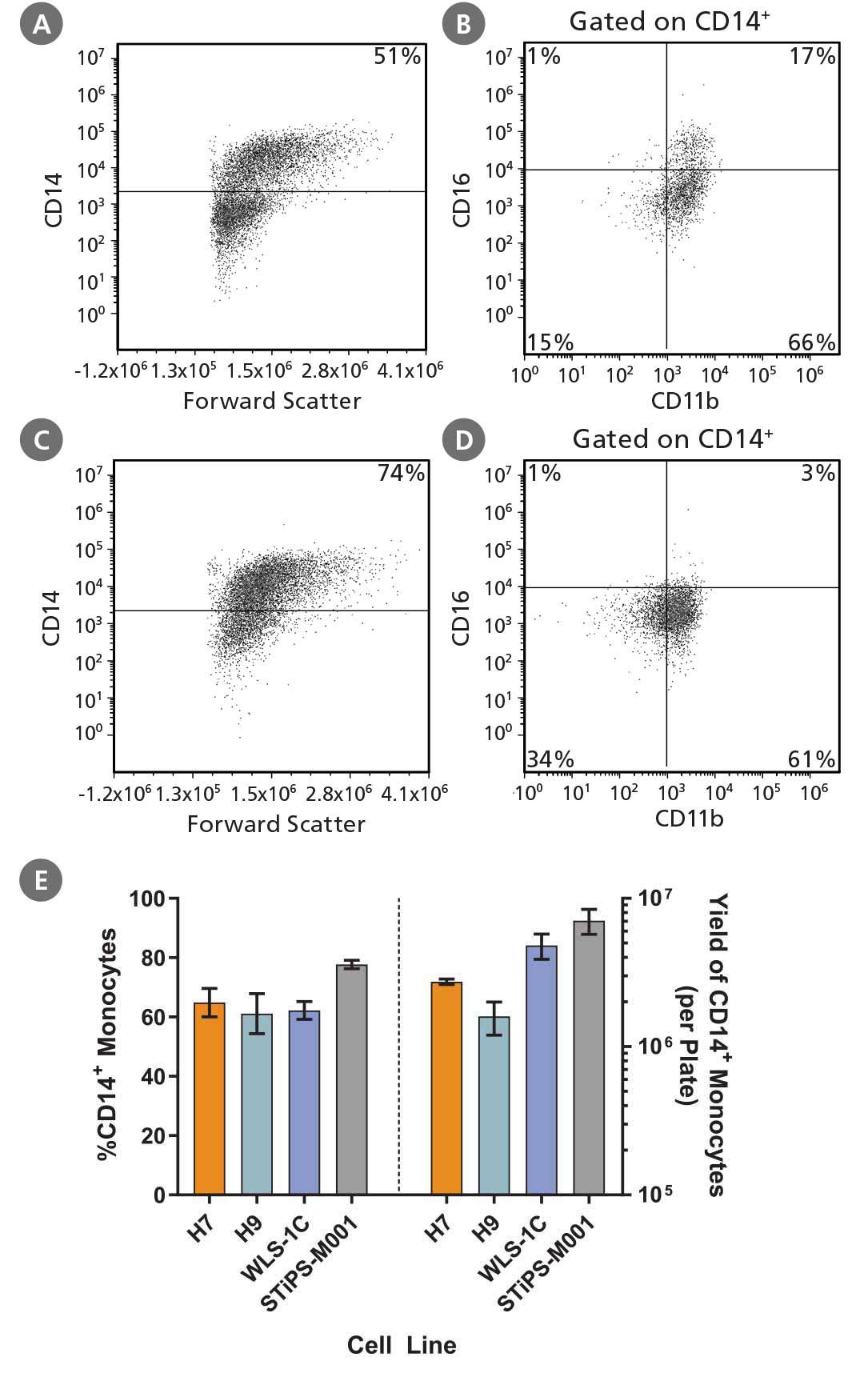
该简便的实验方案在二维贴壁培养中进行。在前 3 天,培养基 A 诱导细胞向中胚层分化。在接下来的 4 天,使用培养基 B 进一步诱导中胚层细胞向造血谱系分化。在第 7 天,将培养基更换为单核细胞分化培养基,以促进其向单核细胞分化。CD14 +单核细胞最早可从第 14 天开始从培养上清液中直接收集,并可在后续培养过程中多次收集。CD14 +细胞的峰值频率通常在 60% - 80%之间。
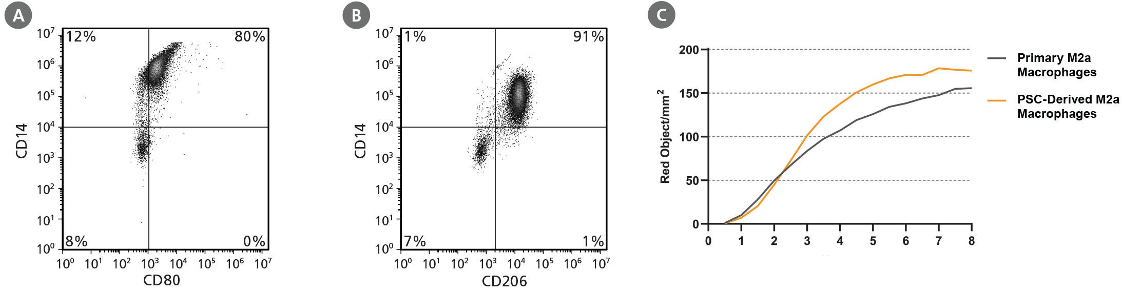
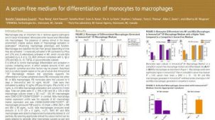
hPSC 衍生的单核细胞可分别使用ImmunoCult™ 树突状细胞培养试剂盒或ImmunoCult™-SF 巨噬细胞培养基进一步分化为树突状细胞或巨噬细胞。
为了方便起见,用于配制单核细胞分化培养基所需的成分StemSpan™ SFEM II和STEMdiff™ 单核细胞分化添加剂 (100X)也可单独购买。
分类
专用培养基
细胞类型
树突状细胞(DCs),巨噬细胞,单核细胞,髓系细胞,多能干细胞
种属
人
应用
细胞培养,分化,扩增
品牌
STEMdiff
研究领域
疾病建模,药物发现和毒性检测,免疫学,干细胞生物学
制剂类别
无血清
请在《产品说明书》中查找相关支持信息和使用说明,或浏览下方更多实验方案。
本产品专为以下研究领域设计,适用于工作流程中的高亮阶段。探索这些工作流程,了解更多我们为各研究领域提供的其他配套产品。
| 物种 | 人 |
|---|---|
| 配方 | 无血清 |
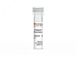
<p>分化为CD14+ 单核细胞的添加物</p>
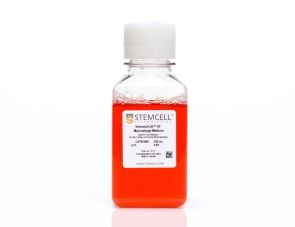
人单核细胞向巨噬细胞分化的无血清培养基
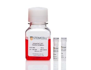
人单核细胞向树突状细胞分化的完整试剂盒
在线联系

 沪公网安备31010102008431号