
 EasySep™小鼠TIL(CD45)正选试剂盒


产品号 #05310_C
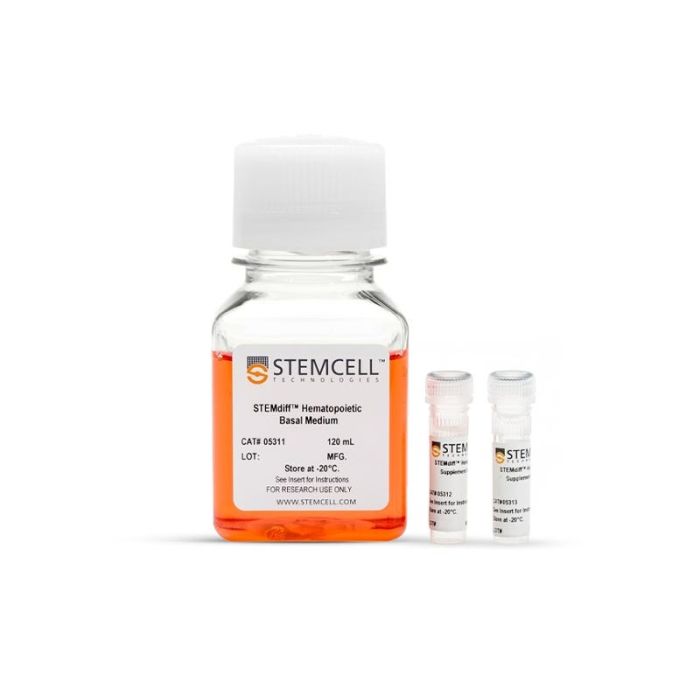
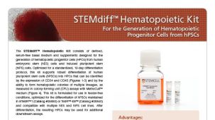
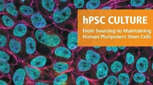
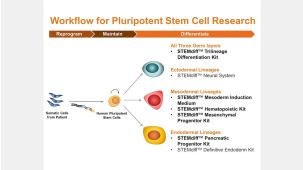
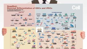
用于将人ES细胞或iPS细胞分化为造血祖细胞
若您需要咨询产品或有任何技术问题,请通过官方电话 400 885 9050 或邮箱 info.cn@stemcell.com 与我们联系。
We developed the STEMdiff™ Hematopoietic Kit so scientists like you can have a reliable source of hPSC-derived hematopoietic progenitor cells for their research.
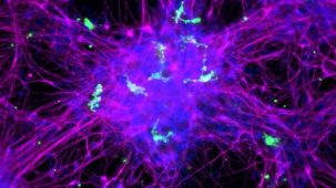
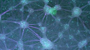
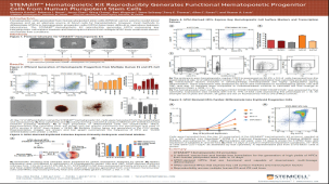
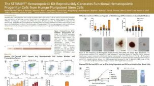
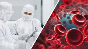
使用 STEMdiff™ 造血试剂盒可从人胚胎干细胞(ES)和诱导多能干细胞(iPS)生成功能性造血祖细胞。该产品采用简便且可重复的培养方案,在无血清、无饲养层的条件下进行分化,可获得表达 CD34、CD45 和 CD43 的造血祖细胞。
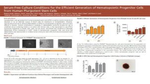
该稳健的两阶段分化方案首先诱导细胞向中胚层分化,随后进一步分化为造血祖细胞。经过 12 天培养后,可获得含有25% 至 65%(平均为 43%)的 CD34⁺CD45⁺ 祖细胞的造血细胞群,其中包括在集落形成单位 (CFU) 测定中能够形成功能性造血集落的祖细胞。
STEMdiff™ 造血试剂盒已针对以下培养基中维持的人多能细胞的分化进行了优化:
• mTeSR™1(目录号 85850)
• mTeSR™ Plus(目录号 100-0276)
• TeSR™-E8™(目录号 05990)
分类
专用培养基
细胞类型
造血干/祖细胞,多能干细胞
种属
人
应用
细胞培养,分化
品牌
STEMdiff
研究领域
干细胞生物学
制剂类别
无血清
请在《产品说明书》中查找相关支持信息和使用说明,或浏览下方更多实验方案。
本产品专为以下研究领域设计,适用于工作流程中的高亮阶段。探索这些工作流程,了解更多我们为各研究领域提供的其他配套产品。
| 物种 | 人 |
|---|---|
| 配方 | 无血清 |
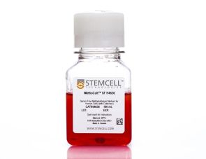
含重组细胞因子的无血清甲基纤维素培养基,适用于人 ES 和 iPS 细胞衍生细胞
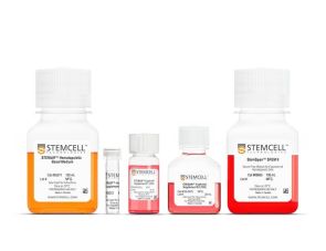
用于将人 ES 细胞或 iPS 细胞分化为红系祖细胞

用于将人胚胎干细胞(ES细胞)或诱导多能干细胞(iPS细胞)分化为巨核细胞和血小板。
<p>cGMP标准、无饲养层的hESC和iPSC维持培养基</p>
在线联系


 沪公网安备31010102008431号