若您需要咨询产品或有任何技术问题,请通过官方电话 400 885 9050 或邮箱 info.cn@stemcell.com 与我们联系。

STEMdiff™ 心肌细胞解离试剂盒

用于解离hPSC衍生的心肌细胞
只有 %1
¥2,838.00

产品号 #(选择产品)

产品号 #05025_C

用于解离hPSC衍生的心肌细胞

产品优势

  • 可保持来源于 hPSC 的心肌细胞完整性,细胞活率超过 80%
  • 提供一种简单且稳定的方法,用于收获和重新铺板 hPSC 衍生的心肌细胞

产品组分包括

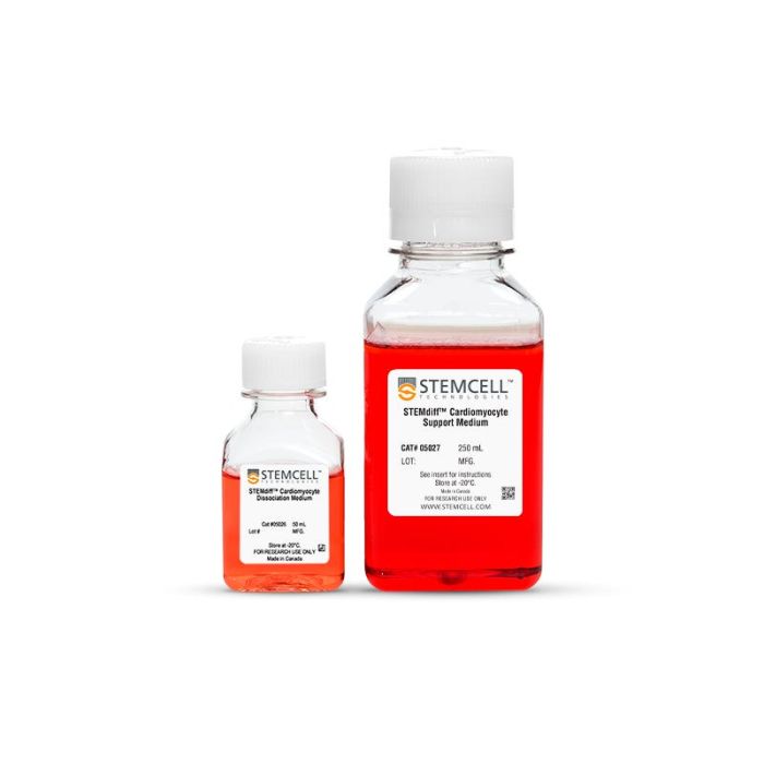
  • STEMdiff™ 心肌细胞解离培养基,50 mL
  • STEMdiff™ 心肌细胞支持培养基,250 mL
专为您的实验方案打造的产品
要查看实验方案所需的所有配套产品,请参阅《实验方案与技术文档》

总览

STEMdiff™ 心肌细胞分离试剂盒包含 STEMdiff™ 心肌细胞分离培养基和 STEMdiff™ 心肌细胞支持培养基。STEMdiff™ 心肌细胞解离培养基可用于收获由人类多能干细胞(hPSCs)分化得到的心肌细胞。这些细胞通过使用 STEMdiff™ 心室心肌细胞分化试剂盒(产品号 #05010)或 STEMdiff™ 心房心肌细胞分化试剂盒(产品号#100-0215)获得,并在 STEMdiff™ 心肌细胞维持培养基(产品号 #05010/#05020)中培养。STEMdiff™ 心肌细胞支持培养基可在收获和重新铺板过程中减少心肌细胞所受的应激,从而保持其活性和功能,为后续应用和分析提供保障。

分类
专用培养基
 
细胞类型
心肌细胞,PSC衍生
 
种属

 
应用
细胞培养
 
品牌
STEMdiff
 
研究领域
干细胞生物学
 

产品说明书及文档

请在《产品说明书》中查找相关支持信息和使用说明,或浏览下方更多实验方案。

Document Type
Product Name
Catalog #
Lot #
Language
Catalog #
05025
Lot #
All
Language
English
Document Type
Safety Data Sheet
Catalog #
05025
Lot #
All
Language
English

应用领域

本产品专为以下研究领域设计,适用于工作流程中的高亮阶段。探索这些工作流程,了解更多我们为各研究领域提供的其他配套产品。

相关材料与文献

技术资料 (5)

文献 (9)

Myosin inhibitor reverses hypertrophic cardiomyopathy in genotypically diverse pediatric iPSC-cardiomyocytes to mirror variant correction C. Kinnear et al. Cell Reports Medicine 2024 Apr

Abstract

Pathogenic variants in MYH7 and MYBPC3 account for the majority of hypertrophic cardiomyopathy (HCM). Targeted drugs like myosin ATPase inhibitors have not been evaluated in children. We generate patient and variant-corrected iPSC-cardiomyocytes (CMs) from pediatric HCM patients harboring single variants in MYH7 ( V606M ; R453C ),MYBPC3 ( G148R ) or digenic variants ( MYBPC3 P955fs,TNNI3 A157V ). We also generate CMs harboring MYBPC3 mono- and biallelic variants using CRISPR editing of a healthy control. Compared with isogenic and healthy controls,variant-positive CMs show sarcomere disorganization,higher contractility,calcium transients,and ATPase activity. However,only MYH7 and biallelic MYBPC3 variant-positive CMs show stronger myosin-actin binding. Targeted myosin ATPase inhibitors show complete rescue of the phenotype in variant-positive CMs and in cardiac Biowires to mirror isogenic controls. The response is superior to verapamil or metoprolol. Myosin inhibitors can be effective in genotypically diverse HCM highlighting the need for myosin inhibitor drug trials in pediatric HCM.
ELM2?SANT Domain?Containing Scaffolding Protein 1 Regulates Differentiation and Maturation of Cardiomyocytes Derived From Human?Induced Pluripotent Stem Cells Journal of the American Heart Association: Cardiovascular and Cerebrovascular Disease 2024 Jun

Abstract

BackgroundELMSAN1 (ELM2?SANT domain?containing scaffolding protein 1) is a newly identified scaffolding protein of the MiDAC (mitotic deacetylase complex),playing a pivotal role in early embryonic development. Studies on Elmsan1 knockout mice showed that its absence results in embryo lethality and heart malformation. However,the precise function of ELMSAN1 in heart development and formation remains elusive. To study its potential role in cardiac lineage,we employed human?induced pluripotent stem cells (hiPSCs) to model early cardiogenesis and investigated the function of ELMSAN1.Methods and ResultsWe generated ELMSAN1?deficient hiPSCs through knockdown and knockout techniques. During cardiac differentiation,ELMSAN1 depletion inhibited pluripotency deactivation,decreased the expression of cardiac?specific markers,and reduced differentiation efficiency. The impaired expression of genes associated with contractile sarcomere structure,calcium handling,and ion channels was also noted in ELMSAN1?deficient cardiomyocytes derived from hiPSCs. Additionally,through a series of structural and functional assessments,we found that ELMSAN1?null hiPSC cardiomyocytes are immature,exhibiting incomplete sarcomere Z?line structure,decreased calcium handling,and impaired electrophysiological properties. Of note,we found that the cardiac?specific role of ELMSAN1 is likely associated with histone H3K27 acetylation level. The transcriptome analysis provided additional insights,indicating maturation reduction with the energy metabolism switch and restored cell proliferation in ELMSAN1 knockout cardiomyocytes.ConclusionsIn this study,we address the significance of the direct involvement of ELMSAN1 in the differentiation and maturation of hiPSC cardiomyocytes. We first report the impact of ELMSAN1 on multiple aspects of hiPSC cardiomyocyte generation,including cardiac differentiation,sarcomere formation,calcium handling,electrophysiological maturation,and proliferation.
P53 Activation Promotes Maturational Characteristics of Pluripotent Stem Cell‐Derived Cardiomyocytes in 3‐Dimensional Suspension Culture Via FOXO‐FOXM1 Regulation N. Velayutham et al. Journal of the American Heart Association: Cardiovascular and Cerebrovascular Disease 2024 Jun

Abstract

Current protocols generate highly pure human induced pluripotent stem cell–derived cardiomyocytes (hiPSC‐CMs) in vitro that recapitulate characteristics of mature in vivo cardiomyocytes. Yet,a risk of arrhythmias exists when hiPSC‐CMs are injected into large animal models. Thus,understanding hiPSC‐CM maturational mechanisms is crucial for clinical translation. Forkhead box (FOX) transcription factors regulate postnatal cardiomyocyte maturation through a balance between FOXO and FOXM1. We also previously demonstrated that p53 activation enhances hiPSC‐CM maturation. Here,we investigate whether p53 activation modulates the FOXO/FOXM1 balance to promote hiPSC‐CM maturation in 3‐dimensional suspension culture. Three‐dimensional cultures of hiPSC‐CMs were treated with Nutlin‐3a (p53 activator,10 μM),LOM612 (FOXO relocator,5 μM),AS1842856 (FOXO inhibitor,1 μM),or RCM‐1 (FOXM1 inhibitor,1 μM),starting 2 days after onset of beating,with dimethyl sulfoxide (0.2% vehicle) as control. P53 activation promoted hiPSC‐CM metabolic and electrophysiological maturation alongside FOXO upregulation and FOXM1 downregulation,in n=3 to 6 per group for all assays. FOXO inhibition significantly decreased expression of cardiac‐specific markers such as TNNT2. In contrast,FOXO activation or FOXM1 inhibition promoted maturational characteristics such as increased contractility,oxygen consumption,and voltage peak maximum upstroke velocity,in n=3 to 6 per group for all assays. Further,by single‐cell RNA sequencing of n=2 LOM612‐treated cells compared with dimethyl sulfoxide,LOM612‐mediated FOXO activation promoted expression of cardiac maturational pathways. We show that p53 activation promotes FOXO and suppresses FOXM1 during 3‐dimensional hiPSC‐CM maturation. These results expand our understanding of hiPSC‐CM maturational mechanisms in a clinically‐relevant 3‐dimensional culture system.

更多信息

更多信息
物种
质量保证:

产品仅供研究使用,不用于针对人或动物的诊断或治疗。 欲获悉更多关于STEMCELL的质控信息,请访问 STEMCELL.CN/COMPLIANCE.
Copyright © 2026 by STEMCELL Technologies. All rights reserved.

在线联系