High NLRC5 Expression Is Associated with an Immunosuppressive Tumor Microenvironment and Poor Prognosis in Esophageal Squamous Cell Carcinoma
Background: Immunotherapy efficacy in esophageal squamous cell carcinoma (ESCC) is often limited by an immunosuppressive tumor microenvironment (TME). NLRC5,a key regulator of MHC-I antigen presentation,exhibits context-dependent roles in tumor immunity; however,its function in ESCC remains unclear. This study aimed to systematically investigate the expression pattern,prognostic value,and immunological role of NLRC5 in ESCC. Methods: An integrated analysis of bulk RNA sequencing and single-cell RNA sequencing (scRNA-seq) data was performed using multiple cohorts,including The Cancer Genome Atlas,Gene Expression Omnibus,and an in-house ESCC cohort. Differential expression,survival analysis,immune infiltration estimation,and functional enrichment analyses were conducted to elucidate the role of NLRC5 in the tumor microenvironment. Results: NLRC5 was significantly upregulated in ESCC and its high expression independently predicted poor patient survival. Although NLRC5 expression was associated with increased CD8+ T cell infiltration,it was paradoxically accompanied by features of T-cell exhaustion and elevated expression of multiple immune checkpoints. Moreover,NLRC5-high tumors were enriched in transcriptional programs related to PANoptosis,indicating an additional immunosuppressive mechanism within the TME. Conclusions: NLRC5 is not only a prognostic biomarker but also a key modulator of an immune-active yet functionally suppressed tumor microenvironment in ESCC. These findings highlight NLRC5 as a potential therapeutic target for restoring effective antitumor immunity.
View Publication
产品号#:
19051
19051RF
产品名:
EasySep™人T细胞富集试剂盒
RoboSep™ 人T细胞富集试剂盒含滤芯吸头
E. Trolio et al. (Mar 2026)
STAR Protocols 7 2
Protocol for immunomagnetic enrichment of T cells from complex murine tissues
SummaryT cells are the central effectors and regulators of the adaptive immune response. This protocol provides a step-by-step approach for isolating and enriching total T cells by negative selection from complex murine tissues,including bone marrow (BM),liver,heart,and kidneys. We describe steps for tissue harvesting,preparation of single-cell suspensions,and immunomagnetic enrichment. We then outline procedures for flow cytometric assessment of cell purity and viability. This protocol enables efficient recovery of high-quality T cells for reliable downstream analyses. Graphical abstract Highlights•Isolation of leukocytes from murine BM,liver,heart and kidneys•Non-enzymatic dissociation of kidney and heart tissue•Protocol for immunomagnetic enrichment of T cells•Flow cytometry analysis of T cell purity and viability Publisher’s note: Undertaking any experimental protocol requires adherence to local institutional guidelines for laboratory safety and ethics. T cells are the central effectors and regulators of the adaptive immune response. This protocol provides a step-by-step approach for isolating and enriching total T cells by negative selection from complex murine tissues,including bone marrow (BM),liver,heart,and kidneys. We describe steps for tissue harvesting,preparation of single-cell suspensions,and immunomagnetic enrichment. We then outline procedures for flow cytometric assessment of cell purity and viability. This protocol enables efficient recovery of high-quality T cells for reliable downstream analyses.
View Publication
产品号#:
18000
20104
20124
产品名:
EasySep™磁极
RoboSep™ 缓冲液
RoboSep™ 缓冲液 (5X浓缩液)
A. Ricafrente et al. (Apr 2026)
PLOS Pathogens 22 4
GM-CSF orchestrates monocyte and granulocyte responses to Cryptococcus gattii
Cryptococcus gattii is an emerging fungal pathogen that is acquired through the respiratory tract and causes invasive infections in both immunocompromised and otherwise healthy people. Many of these apparently immunocompetent patients are subsequently found to have autoantibodies against the pleiotropic cytokine GM-CSF. In this study,we investigated the potential role of GM-CSF (or CSF2) in the host response to C. gattii using a murine model of infection. Interestingly,infected Csf2-/- mice were found to have significantly improved survival and decreased lung fungal burden compared to wild type (WT) mice. We determined that during C. gattii infection,GM-CSF promotes the differentiation of monocytes into alveolar and interstitial macrophages. When these macrophages are ablated in CCR2-DTR+ mice,there is a corresponding improvement in survival with decreased lung fungal burden,thus phenocopying Csf2-/- mice. WT bone-marrow derived macrophages challenged with C. gattii and interstitial and alveolar macrophages from infected WT mice are unable to undergo M1 polarization,suggesting that monocyte-derived macrophages (moMacs) are rendered permissive for fungal proliferation. Therefore,GM-CSF and moMacs mediate immune responses that are harmful to the host. We further found that GM-CSF and moMacs preferentially promote the influx of eosinophils over neutrophils into the infected lung which is associated with substantial inflammatory lung pathology. Ablation of neutrophils using Mrp8cretg iDTR+ mice significantly increased C. gattii burden in the lungs,indicating that GM-CSF and moMacs block the entry of these beneficial,fungal-clearing granulocytes during infection. Altogether,our results show that GM-CSF plays a key role in impeding host anti-fungal responses to C. gattii by coordinating monocyte-derived macrophages and granulocyte activity and crosstalk. Author summaryCryptococcus gattii is an environmental fungus that can cause severe lung and brain infections after inhalation through the respiratory tract. C. gattii causes disease in patients with known immune deficits but also in individuals that are apparently healthy. Studies on otherwise healthy people who become infected with C. gattii suggest that they may have a previously unrecognized problem involving granulocyte macrophage-colony stimulating factor (GM-CSF),a cytokine,or messenger protein,that is an important part of the immune system. Here,we investigate the role of GM-CSF in the immune response to C. gattii using a mouse model of infection. We find that C. gattii increases GM-CSF in the lungs,leading to the influx of immune cells,including monocyte-derived macrophages and eosinophils,while inhibiting the entry of neutrophils. The macrophages and eosinophils allow the fungus to proliferate and cause inflammatory damage to the lungs,which is ultimately fatal. The absence of neutrophils also contributes to fungal growth,as these immune cells would otherwise be able to help kill the fungus. Our study provides new insight into how GM-CSF regulates immunity to C. gattii and has important implications as to the mechanisms that govern susceptibility to this infection.
View Publication
产品号#:
100-0659
产品名:
EasySep™ 小鼠F4/80正选试剂盒
M. F. Sentmanat et al. (Apr 2026)
Nucleic Acids Research 54 6
Efficient multi-kilobase knock-ins in mice and cell lines using CRISPR/Cas9 and rAAV donors with unbiased whole-genome characterization by LOCK-seq
Multi-kilobase knock-ins (KIs) are a necessary,yet challenging type of genome editing to create and characterize in cell lines and animals. The combination of rAAV donor transduction and electroporation of single-cell mouse embryos with Cas9/gRNA ribonucleoprotein complex enables highly efficient KI,but the insert size is limited by the viral packaging capacity. Here,we report the creation of up to 6.7 kb precise KI achieved in one step by using three rAAVs designed to insert one after the other. To fully characterize the edited genome with large KIs,we developed LOCK-seq (LOng-read sequencing of Captured Kilo-base targets),where relevant genomic regions are enriched via hybridization,achieving over 100-fold greater coverage compared with other long-read methods with enrichment. LOCK-seq simultaneously detects the presence of precise KI alleles,imprecision in the insert and donor concatenation,genotypes of non-KI alleles,and more importantly,uniquely identifies and localizes random integration of the full or partial donor(s). Additionally,the multi-rAAV donor approach is successfully applied to cell lines,including lines intolerant of plasmid DNA,whereas LOCK-seq reliably and efficiently screens for KI clones. Together,the two approaches significantly improve the creation and precision of knock-in models.
View Publication
产品号#:
100-0276
100-0483
100-0484
100-1130
产品名:
mTeSR™ Plus
Hausser Scientificᵀᴹ 明线血球计数板
ReLeSR™
mTeSR™ Plus
T. Shibata et al. (Apr 2026)
Signal Transduction and Targeted Therapy 11
Bioengineered iPSC-derived human macrophages with increased angiotensin-converting enzyme (ACE) expression suppress solid tumor growth
The potential of the immune system to decrease cancer progression is widely recognized and has led to the development of innovative anti-cancer immunotherapies. Here,we studied human macrophages derived from genetically engineered iPSCs (iMac) with angiotensin-converting enzyme (ACE) expression regulatable by a doxycycline (dox)-inducible promoter as a novel anti-cancer immunotherapy. Increased ACE expression in iMac (cells now termed ACE-iMac) augments polarization towards an M1 macrophage phenotype characterized by increased production of proinflammatory cytokines,reactive oxygen species,nitric oxide,and an RNA profile indicating an aggressive immune response. ACE-iMac kills tumor cells in vitro significantly better than iMac. In vivo,studies using tumor xenografts for melanoma,breast cancer,and head and neck squamous cell carcinoma (HNSCC) showed a highly significant 3.4- to 7.2-fold reduction in solid tumor size following ACE-expressing ACE-iMac immunotherapy as compared to results with iMac. To further investigate the impact of ACE on human anti-tumor responses,we developed a humanized BLT-NSG mouse model with a fully functional adaptive immune system. Here,ACE-iMac treatment significantly reduced the growth of human melanoma xenografts by enhancing the activation of human T cells and NK cells. In conclusion,enhancing ACE expression in human-derived macrophages (ACE-iMac) greatly amplifies their anti-cancer phenotype,offering a compelling new therapeutic strategy with the potential to improve clinical outcomes for cancer patients.
View Publication
产品号#:
100-0276
100-1130
产品名:
mTeSR™ Plus
mTeSR™ Plus
K. T. Wagner et al. (Apr 2026)
APL Bioengineering 10 2
Mapping the miRNA landscape of primitive macrophage extracellular vesicles highlights their pro-vasculogenic effects in engineered human cardiac tissue
Resident cardiac macrophages,derived from primitive yolk sac precursors during embryogenesis,have increasingly been recognized for their distinct phenotype and functions in regulating homeostasis of the human heart. However,the profile of their extracellular vesicles (EVs) in cardiac signaling and regulation remains uncharted. Here,we employ differentiation of human pluripotent stem cell-derived primitive macrophages (Mac),harvesting their secreted EVs and performing in-depth characterization of associated microRNAs (miRNAs). Primitive macrophages secreted nanoscale EVs that expressed canonical EV markers,and miRNA sequencing highlighted a diverse and unique profile of miRNAs when compared to EVs sourced from other principal cardiac cell lineages and published data from monocyte-derived cells. In particular,we noted the abundance and enrichment of vascular-modulatory let-7 miRNAs and miR-126-3p. Functional screening of Mac-EVs in a 3D model of in vitro cardiac vasculogenesis confirmed enhanced early endothelial cell organization and branching. Establishing a reference for the human Mac-EV miRNome enables further hypothesis-driven mechanistic tests of Mac-EV miRNAs in mediating cardiac physiology and disease,opening the door to identification of therapeutic targets and modalities for cardiac repair.
View Publication

 EasySep™小鼠TIL(CD45)正选试剂盒


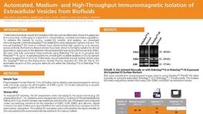 科学海报Automated, Medium- and High-Throughput Immunomagnetic Isolation of Extracellular Vesicles from Biofluids

 沪公网安备31010102008431号