
 EasySep™小鼠TIL(CD45)正选试剂盒


技术资料
-
技术窍门人肠道类器官培养的常见问题
-
产品说明书10000000383-PIS_02.pdf
产品号#:
07940
07931
07930
07955
07959
07952
100-1061
产品名:
CryoStor® CS10
CryoStor® CS10
CryoStor® CS10
CryoStor® CS10
CryoStor® CS10
CryoStor® CS10
CryoStor® CS10
-
-
安全数据表DX21420-SDS_2_1_0.pdf
产品号#:
19555
19555RF
产品名:
EasySep™人Naïve CD4+ T细胞分选试剂盒
RoboSep™ 人Naïve CD4+ T细胞分选试剂盒
-
-
-
-
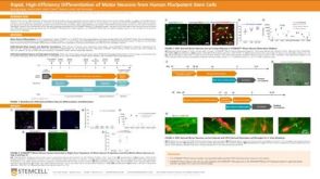 科学海报Rapid, High-Efficiency Differentiation of Motor Neurons from Human Pluripotent Stem Cells
Conference:
FENS 2022
-
-
-

 沪公网安备31010102008431号