Li W et al. (JAN 2009)
The Journal of biological chemistry 284 1 218--28
The serine protease marapsin is expressed in stratified squamous epithelia and is up-regulated in the hyperproliferative epidermis of psoriasis and regenerating wounds.
The trypsin-like serine protease marapsin is a member of the large protease gene cluster at human chromosome 16p13.3,which also contains the structurally related proteases testisin,tryptase epsilon,tryptase gamma,and EOS. To gain insight into the biological functions of marapsin,we undertook a detailed gene expression analysis. It showed that marapsin expression was restricted to tissues containing stratified squamous epithelia and was absent or only weakly expressed in all other tissues,including the pancreas. Marapsin was constitutively expressed in nonkeratinizing stratified squamous epithelia of human esophagus,tonsil,cervix,larynx,and cornea. In the keratinizing stratified squamous epidermis of skin,however,its expression was induced only during epidermal hyperproliferation,such as in psoriasis and in murine wound healing. In fact,marapsin was the second most strongly up-regulated protease in psoriatic lesions,where expression was localized to the upper region of the hyperplastic epidermis. Similarly,in the hyperproliferative epithelium of regenerating murine skin wounds,marapsin localized to the suprabasal layers,where keratinocytes undergo squamous differentiation. The transient up-regulation of marapsin,which closely correlated with re-epithelialization,was virtually absent in a genetic mouse model of delayed wound closure. These results suggested a function during the process of re-epithelialization. Furthermore,in reconstituted human epidermis,a model system of epidermal differentiation,members of the IL-20 subfamily of cytokines,such as IL-22,induced marapsin expression. Consistent with a physiologic role in marapsin regulation,IL-22 was also strongly expressed in re-epithelializing skin wounds. Marapsin's restricted expression,localization,and cytokine-inducible expression suggest a role in the terminal differentiation of keratinocytes in hyperproliferating squamous epithelia.
View Publication
产品号#:
03800
03801
03802
03803
03804
03805
03806
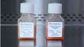
产品名:
ClonaCell™-HY杂交瘤试剂盒
ClonaCell™-HY培养基A
ClonaCell™-HY 培养基 B
ClonaCell™-HY 培养基 C
ClonaCell™-HY 培养基 D
ClonaCell™-HY 培养基 E
ClonaCell™-HY PEG
Ling SSM et al. (JUN 2015)
PLOS ONE 10 6 e0131460
Instrumental Role of Helicobacter pylori γ-Glutamyl Transpeptidase in VacA-Dependent Vacuolation in Gastric Epithelial Cells
Helicobacter pylori causes cellular vacuolation in host cells,a cytotoxic event attributed to vacuolating cytotoxin (VacA) and the presence of permeant weak bases such as ammonia. We report here the role of γ-glutamyl transpeptidase (GGT),a constitutively expressed secretory enzyme of H. pylori,in potentiating VacA-dependent vacuolation formation in H. pylori-infected AGS and primary gastric cells. The enhancement is brought about by GGT hydrolysing glutamine present in the extracellular medium,thereby releasing ammonia which accentuates the VacA-induced vacuolation. The events of vacuolation in H. pylori wild type (WT)- and Δggt-infected AGS cells were first captured and visualized by real-time phase-contrast microscopy where WT was observed to induce more vacuoles than Δggt. By using semi-quantitative neutral red uptake assay,we next showed that Δggt induced significantly less vacuolation in AGS and primary gastric epithelial cells as compared to the parental strain (Ptextless0.05) indicating that GGT potentiates the vacuolating effect of VacA. Notably,vacuolation induced by WT was significantly reduced in the absence of GGT substrate,glutamine (Ptextless0.05) or in the presence of a competitive GGT inhibitor,serine-borate complex. Furthermore,the vacuolating ability of Δggt was markedly restored when co-incubated with purified recombinant GGT (rGGT),although rGGT itself did not induce vacuolation independently. Similarly,the addition of exogenous ammonium chloride as a source of ammonia also rescued the ability of Δggt to induce vacuolation. Additionally,we also show that monoclonal antibodies against GGT effectively inhibited GGT activity and successfully suppressed H. pylori-induced vacuolation. Collectively,our results clearly demonstrate that generation of ammonia by GGT through glutamine hydrolysis is responsible for enhancing VacA-dependent vacuolation. Our findings provide a new perspective on GGT as an important virulence factor and a promising target in the management of H. pylori-associated gastric diseases.
View Publication
Vieillard V et al. (AUG 2005)
Proceedings of the National Academy of Sciences 102 31 10981--86
NK cytotoxicity against CD4+ T cells during HIV-1 infection: A gp41 peptide induces the expression of an NKp44 ligand
HIV infection leads to a state of chronic immune activation and progressive deterioration in immune function,manifested most recognizably by the progressive depletion of CD4+ T cells. A substantial percentage of natural killer (NK) cells from patients with HIV infection are activated and express the natural cytotoxicity receptor (NCR) NKp44. Here we show that a cellular ligand for NKp44 (NKp44L) is expressed during HIV-1 infection and is correlated with both the progression of CD4+ T cell depletion and the increase of viral load. CD4+ T cells expressing this ligand are highly sensitive to the NK lysis activity mediated by NKp44+ NK cells. The expression of NKp44L is induced by the linear motif NH2-SWSNKS-COOH of the HIV-1 envelope gp41 protein. This highly conserved motif appears critical to the sharp increase in NK lysis of CD4+ T cells from HIV-infected patients. These studies strongly suggest that induction of NKp44L plays a key role in the lysis of CD4+ T cells by activated NK cells in HIV infection and consequently provide a framework for considering how HIV-1 may use NK cell immune surveillance to trigger CD4+ T cells. Understanding this mechanism may help to develop future therapeutic strategies and vaccines against HIV-1 infection.
View Publication
产品号#:
03800
03801
03802
03803
03804
03805
03806
05150
15021
15061
产品名:
ClonaCell™-HY杂交瘤试剂盒
ClonaCell™-HY培养基A
ClonaCell™-HY 培养基 B
ClonaCell™-HY 培养基 C
ClonaCell™-HY 培养基 D
ClonaCell™-HY 培养基 E
ClonaCell™-HY PEG
MyeloCult™ H5100
RosetteSep™人T细胞富集抗体混合物
RosetteSep™人T细胞富集抗体混合物
Matsumoto SC et al. (JAN 2006)
The FASEB Journal 20 3 550--2
Retinal dysfunction in patients with chronic Chagas' disease is associated to anti-Trypanosoma cruzi antibodies that cross-react with rhodopsin
To investigate retinal involvement in chronic Chagas' disease,we performed electroretinography and retinal fluorescein angiography studies in chagasic patients. Our results demonstrated a dissociated electrophysiological response characterized by both an abnormal reduction of the electroretinographic b-wave amplitude and a delayed latency,under the dark-adaptated condition. These alterations are compatible with a selective dysfunction of the rods. Antibodies raised against Trypanosoma cruzi that also interact with beta1-adrenergic receptor blocked light stimulation of cGMP-phosphodiesterase in bovine rod membranes. The specificity from the antibody-rhodopsin interaction was confirmed by Western blot analysis and antigenic competition experiments. Our results suggest an immunomediated rhodopsin blockade. T. cruzi infection probably induces an autoimmune response against rhodopsin in the chronic phase of Chagas' disease through a molecular mimicry mechanism similar to that described previously on cardiac human beta1-adrenergic and M2-cholinergic receptors,all related to the same subfamily of G-protein-coupled receptors.
View Publication

 EasySep™小鼠TIL(CD45)正选试剂盒


 沪公网安备31010102008431号