Physiological β-catenin signaling controls self-renewal networks and generation of stem-like cells from nasopharyngeal carcinoma.
BACKGROUND: A few reports suggested that low levels of Wnt signaling might drive cell reprogramming,but these studies could not establish a clear relationship between Wnt signaling and self-renewal networks. There are ongoing debates as to whether and how the Wnt/β-catenin signaling is involved in the control of pluripotency gene networks. Additionally,whether physiological β-catenin signaling generates stem-like cells through interactions with other pathways is as yet unclear. The nasopharyngeal carcinoma HONE1 cells have low expression of β-catenin and wild-type expression of p53,which provided a possibility to study regulatory mechanism of stemness networks induced by physiological levels of Wnt signaling in these cells.backslashnbackslashnRESULTS: Introduction of increased β-catenin signaling,haploid expression of β-catenin under control by its natural regulators in transferred chromosome 3,resulted in activation of Wnt/β-catenin networks and dedifferentiation in HONE1 hybrid cell lines,but not in esophageal carcinoma SLMT1 hybrid cells that had high levels of endogenous β-catenin expression. HONE1 hybrid cells displayed stem cell-like properties,including enhancement of CD24(+) and CD44(+) populations and generation of spheres that were not observed in parental HONE1 cells. Signaling cascades were detected in HONE1 hybrid cells,including activation of p53- and RB1-mediated tumor suppressor pathways,up-regulation of Nanog-,Oct4-,Sox2-,and Klf4-mediated pluripotency networks,and altered E-cadherin expression in both in vitro and in vivo assays. qPCR array analyses further revealed interactions of physiological Wnt/β-catenin signaling with other pathways such as epithelial-mesenchymal transition,TGF-β,Activin,BMPR,FGFR2,and LIFR- and IL6ST-mediated cell self-renewal networks. Using β-catenin shRNA inhibitory assays,a dominant role for β-catenin in these cellular network activities was observed. The expression of cell surface markers such as CD9,CD24,CD44,CD90,and CD133 in generated spheres was progressively up-regulated compared to HONE1 hybrid cells. Thirty-four up-regulated components of the Wnt pathway were identified in these spheres.backslashnbackslashnCONCLUSIONS: Wnt/β-catenin signaling regulates self-renewal networks and plays a central role in the control of pluripotency genes,tumor suppressive pathways and expression of cancer stem cell markers. This current study provides a novel platform to investigate the interaction of physiological Wnt/β-catenin signaling with stemness transition networks.
View Publication
Thirukkumaran CM et al. (JUL 2003)
Blood 102 1 377--87
Reovirus oncolysis as a novel purging strategy for autologous stem cell transplantation.
Hematologic stem cell rescue after high-dose cytotoxic therapy is extensively used for the treatment of many hematopoietic and solid cancers. Gene marking studies suggest that occult tumor cells within the autograft may contribute to clinical relapse. To date purging of autografts contaminated with cancer cells has been unsuccessful. The selective oncolytic property of reovirus against myriad malignant histologies in in vitro,in vivo,and ex vivo systems has been previously demonstrated. In the present study we have shown that reovirus can successfully purge cancer cells within autografts. Human monocytic and myeloma cell lines as well as enriched ex vivo lymphoma,myeloma,and Waldenström macroglobulinemia patient tumor specimens were used in an experimental purging model. Viability of the cell lines or purified ex vivo tumor cells of diffuse large B-cell lymphoma,chronic lymphocytic leukemia,Waldenström macroglobulinemia,and small lymphocytic lymphoma was significantly reduced after reovirus treatment. Further,[35S]-methionine labeling and sodium dodecyl sulfate-polyacrylamide gel electrophoresis (SDS-PAGE) of cellular proteins demonstrated reovirus protein synthesis and disruption of host cell protein synthesis as early as 24 hours. Admixtures of apheresis product with the abovementioned tumor cells and cell lines treated with reovirus showed complete purging of disease. In contrast,reovirus purging of enriched ex vivo multiple myeloma,Burkitt lymphoma,and follicular lymphoma was incomplete. The oncolytic action of reovirus did not affect CD34+ stem cells or their long-term colony-forming assays even after granulocyte colony-stimulating factor (G-CSF) stimulation. Our results indicate the ex vivo use of an unattenuated oncolytic virus as an attractive purging strategy for autologous stem cell transplantations.
View Publication
产品号#:
04434
04444
09600
09650
84434
84444
产品名:
MethoCult™ H4434 Classic
MethoCult™ H4434 Classic
StemSpan™ SFEM
StemSpan™ SFEM
Coffman KT et al. (NOV 2003)
Cancer Research 63 22 7907--12
Differential EphA2 epitope display on normal versus malignant cells.
The EphA2 receptor tyrosine kinase is overexpressed in many different types of human cancers where it functions as a powerful oncoprotein. Dramatic changes in the subcellular localization and function of EphA2 have also been linked with cancer,and in particular,unstable cancer cell-cell contacts prevent EphA2 from stably binding its ligand on the surface of adjoining cells. This change is important in light of evidence that ligand binding causes EphA2 to transmit signals that negatively regulate tumor cell growth and invasiveness and also induce EphA2 degradation. On the basis of these properties,we have begun to target EphA2 on tumor cells using agonistic antibodies,which mimic the consequences of ligand binding. In our present study,we show that a subset of agonistic EphA2 antibodies selectively bind epitopes on malignant cells,which are not available on nontransformed epithelial cells. We also show that such epitopes arise from differential cell-cell adhesions and that the stable intercellular junctions of nontransformed epithelial cells occlude the binding site for ligand,as well as this subset of EphA2 antibodies. Finally,we demonstrate that antibody targeting of EphA2 decreases tumor cell growth as measured using xenograft tumor models and found that the mechanism of antibody action relates to EphA2 protein degradation in vivo. Taken together,these results suggest new opportunities for therapeutic targeting of the large number of different cancers that express EphA2 in a manner that could minimize potential toxicities to normal cells.
View Publication
产品号#:
03800
03801
03802
03803
03804
03805
03806
产品名:
ClonaCell™-HY杂交瘤试剂盒
ClonaCell™-HY培养基A
ClonaCell™-HY 培养基 B
ClonaCell™-HY 培养基 C
ClonaCell™-HY 培养基 D
ClonaCell™-HY 培养基 E
ClonaCell™-HY PEG
Esplugues E et al. (JUN 2005)
Blood 105 11 4399--406
Induction of tumor NK-cell immunity by anti-CD69 antibody therapy.
The leukocyte activation marker CD69 is a novel regulator of the immune response,modulating the production of cytokines including transforming growth factor-beta (TGF-beta). We have generated an antimurine CD69 monoclonal antibody (mAb),CD69.2.2,which down-regulates CD69 expression in vivo but does not deplete CD69-expressing cells. Therapeutic administration of CD69.2.2 to wild-type mice induces significant natural killer (NK) cell-dependent antitumor responses to major histocompatibility complex (MHC) class I low RMA-S lymphomas and to RM-1 prostatic carcinoma lung metastases. These in vivo antitumor responses are comparable to those seen in CD69(-/-) mice. Enhanced host NK cytotoxic activity correlates with a reduction in NK-cell TGF-beta production and is independent of tumor priming. In vitro studies demonstrate the novel ability of anti-CD69 mAbs to activate resting NK cells in an Fc receptor-independent manner,resulting in a substantial increase in both NK-cell cytolytic activity and interferon gamma (IFNgamma) production. Modulation of the innate immune system with monoclonal antibodies to host CD69 thus provides a novel means to antagonize tumor growth and metastasis.
View Publication
产品号#:
09600
09650
产品名:
StemSpan™ SFEM
StemSpan™ SFEM
Wittman VP et al. (SEP 2006)
The Journal of Immunology 177 6 4187--95
Antibody targeting to acClass I MHC-peptide epitope promotes tumor cell death
Therapeutic mAbs that target tumor-associated Ags on the surface of malignant cells have proven to be an effective and specific option for the treatment of certain cancers. However,many of these protein markers of carcinogenesis are not expressed on the cells' surface. Instead these tumor-associated Ags are processed into peptides that are presented at the cell surface,in the context of MHC class I molecules,where they become targets for T cells. To tap this vast source of tumor Ags,we generated a murine IgG2a mAb,3.2G1,endowed with TCR-like binding specificity for peptide-HLA-A*0201 (HLA-A2) complex and designated this class of Ab as TCR mimics (TCRm). The 3.2G1 TCRm recognizes the GVL peptide (GVLPALPQV) from human chorionic gonadotropin beta presented by the peptide-HLA-A*0201 complex. When used in immunofluorescent staining reactions using GVL peptide-loaded T2 cells,the 3.2G1 TCRm specifically stained the cells in a peptide and Ab concentration-dependent manner. Staining intensity correlated with the extent of cell lysis by complement-dependent cytotoxicity (CDC),and a peptide concentration-dependent threshold level existed for the CDC reaction. Staining of human tumor lines demonstrated that 3.2G1 TCRm was able to recognize endogenously processed peptide and that the breast cancer cell line MDA-MB-231 highly expressed the target epitope. The 3.2G1 TCRm-mediated CDC and Ab-dependent cellular cytotoxicity of a human breast carcinoma line in vitro and inhibited in vivo tumor implantation and growth in nude mice. These results provide validation for the development of novel TCRm therapeutic reagents that specifically target and kill tumors via recognition and binding to MHC-peptide epitopes.
View Publication
Altered B-cell receptor signaling kinetics distinguish human follicular lymphoma B cells from tumor-infiltrating nonmalignant B cells.
The B-cell receptor (BCR) transmits life and death signals throughout B-cell development,and altered BCR signaling may be required for survival of B-lymphoma cells. We used single-cell signaling profiles to compare follicular lymphoma (FL) B cells and nonmalignant host B cells within individual patient biopsies and identified BCR-mediated signaling events specific to lymphoma B cells. Expression of CD20,Bcl-2,and BCR light chain isotype (kappa or lambda) distinguished FL tumor B-cell and nontumor host B-cell subsets within FL patient biopsies. BCR-mediated signaling via phosphorylation of Btk,Syk,Erk1/2,and p38 occurred more rapidly in tumor B cells from FL samples than in infiltrating nontumor B cells,achieved greater levels of per-cell signaling,and sustained this level of signaling for hours longer than nontumor B cells. The timing and magnitude of BCR-mediated signaling in nontumor B cells within an FL sample instead resembled that observed in mature B cells from the peripheral blood of healthy subjects. BCR signaling pathways that are potentiated specifically in lymphoma cells should provide new targets for therapeutic attention.
View Publication
产品号#:
09850
产品名:
Tripp A et al. (NOV 2005)
Journal of virology 79 22 14069--78
Induction of cell cycle arrest by human T-cell lymphotropic virus type 1 Tax in hematopoietic progenitor (CD34+) cells: modulation of p21cip1/waf1 and p27kip1 expression.
Human T-cell lymphotropic virus type 1 (HTLV-1) is the etiologic agent of adult T-cell leukemia,an aggressive CD4(+) malignancy. Although HTLV-2 is highly homologous to HTLV-1,infection with HTLV-2 has not been associated with lymphoproliferative disorders. Lentivirus-mediated transduction of CD34(+) cells with HTLV-1 Tax (Tax1) induced G(0)/G(1) cell cycle arrest and resulted in the concomitant suppression of multilineage hematopoiesis in vitro. Tax1 induced transcriptional upregulation of the cdk inhibitors p21(cip1/waf1) (p21) and p27(kip1) (p27),and marked suppression of hematopoiesis in immature (CD34(+)/CD38(-)) hematopoietic progenitor cells in comparison to CD34(+)/CD38(+) cells. HTLV-1 infection of CD34(+) cells also induced p21 and p27 expression. Tax1 also protected CD34(+) cells from serum withdrawal-mediated apoptosis. In contrast,HTLV-2 Tax (Tax2) did not detectably alter p21 or p27 gene expression,failed to induce cell cycle arrest,failed to suppress hematopoiesis in CD34(+) cells,and did not protect cells from programmed cell death. A Tax2/Tax1 chimera encoding the C-terminal 53 amino acids of Tax1 fused to Tax2 (Tax(221)) displayed a phenotype in CD34(+) cells similar to that of Tax1,suggesting that unique domains encoded within the C terminus of Tax1 may account for the phenotypes displayed in human hematopoietic progenitor cells. These remarkable differences in the activities of Tax1 and Tax2 in CD34(+) hematopoietic progenitor cells may underlie the sharp differences observed in the pathogenesis resulting from infection with HTLV-1 and HTLV-2.
View Publication

 EasySep™小鼠TIL(CD45)正选试剂盒


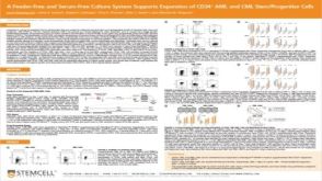 科学海报A Feeder-Free and Serum-Free Culture System Supports Expansion of CD34+ AML and CML Stem/Progenitor Cells


 沪公网安备31010102008431号