Physiological β-catenin signaling controls self-renewal networks and generation of stem-like cells from nasopharyngeal carcinoma.
BACKGROUND: A few reports suggested that low levels of Wnt signaling might drive cell reprogramming,but these studies could not establish a clear relationship between Wnt signaling and self-renewal networks. There are ongoing debates as to whether and how the Wnt/β-catenin signaling is involved in the control of pluripotency gene networks. Additionally,whether physiological β-catenin signaling generates stem-like cells through interactions with other pathways is as yet unclear. The nasopharyngeal carcinoma HONE1 cells have low expression of β-catenin and wild-type expression of p53,which provided a possibility to study regulatory mechanism of stemness networks induced by physiological levels of Wnt signaling in these cells.backslashnbackslashnRESULTS: Introduction of increased β-catenin signaling,haploid expression of β-catenin under control by its natural regulators in transferred chromosome 3,resulted in activation of Wnt/β-catenin networks and dedifferentiation in HONE1 hybrid cell lines,but not in esophageal carcinoma SLMT1 hybrid cells that had high levels of endogenous β-catenin expression. HONE1 hybrid cells displayed stem cell-like properties,including enhancement of CD24(+) and CD44(+) populations and generation of spheres that were not observed in parental HONE1 cells. Signaling cascades were detected in HONE1 hybrid cells,including activation of p53- and RB1-mediated tumor suppressor pathways,up-regulation of Nanog-,Oct4-,Sox2-,and Klf4-mediated pluripotency networks,and altered E-cadherin expression in both in vitro and in vivo assays. qPCR array analyses further revealed interactions of physiological Wnt/β-catenin signaling with other pathways such as epithelial-mesenchymal transition,TGF-β,Activin,BMPR,FGFR2,and LIFR- and IL6ST-mediated cell self-renewal networks. Using β-catenin shRNA inhibitory assays,a dominant role for β-catenin in these cellular network activities was observed. The expression of cell surface markers such as CD9,CD24,CD44,CD90,and CD133 in generated spheres was progressively up-regulated compared to HONE1 hybrid cells. Thirty-four up-regulated components of the Wnt pathway were identified in these spheres.backslashnbackslashnCONCLUSIONS: Wnt/β-catenin signaling regulates self-renewal networks and plays a central role in the control of pluripotency genes,tumor suppressive pathways and expression of cancer stem cell markers. This current study provides a novel platform to investigate the interaction of physiological Wnt/β-catenin signaling with stemness transition networks.
View Publication
Rush SZ et al. (AUG 2010)
Neuro-oncology 12 8 790--8
Activation of the Hedgehog pathway in pilocytic astrocytomas.
Pilocytic astrocytoma is commonly viewed as a benign lesion. However,disease onset is most prevalent in the first two decades of life,and children are often left with residual or recurrent disease and significant morbidity. The Hedgehog (Hh) pathway regulates the growth of higher WHO grade gliomas,and in this study,we have evaluated the activation and operational status of this regulatory pathway in pilocytic astrocytomas. Expression levels of the Hh pathway transcriptional target PTCH were elevated in 45% of tumor specimens analyzed (ages 1-22 years) and correlated inversely with patient age. Evaluation of a tissue array revealed oligodendroglioma-like features,pilomyxoid features,infiltration,and necrosis more commonly in specimens from younger patients (below the median patient age of 10 years). Immunohistochemical staining for the Hh pathway components PTCH and GLI1 and the proliferation marker Ki67 demonstrated that patients diagnosed before the age of 10 had higher staining indices than those diagnosed after the age of 10. A significant correlation between Ki67 and PTCH and GLI1 staining indices was measured,and 86% of Ki67-positive cells also expressed PTCH. The operational status of the Hh pathway was confirmed in primary cell culture and could be modulated in a manner consistent with a ligand-dependent mechanism. Taken together,these findings suggest that Hh pathway activation is common in pediatric pilocytic astrocytomas and may be associated with younger age at diagnosis and tumor growth.
View Publication
产品号#:
05751
产品名:
NeuroCult™ NS-A 扩增试剂盒(人)
Liu S and Wicha MS (SEP 2010)
Journal of clinical oncology : official journal of the American Society of Clinical Oncology 28 25 4006--12
Targeting breast cancer stem cells.
There is increasing evidence that many cancers,including breast cancer,contain populations of cells that display stem-cell properties. These breast cancer stem cells,by virtue of their relative resistance to radiation and cytotoxic chemotherapy,may contribute to treatment resistance and relapse. The elucidation of pathways that regulate these cells has led to the identification of potential therapeutic targets. A number of agents capable of targeting breast cancer stem cells in preclinical models are currently entering clinical trials. Assessment of the efficacy of the agents will require development of innovative clinical trial designs with appropriate biologic and clinical end points. The effective targeting of breast cancer stem cells has the potential to significantly improve outcome for women with both early-stage and advanced breast cancer.
View Publication
产品号#:
01700
01705
01702
产品名:
ALDEFLUOR™ 试剂盒
ALDEFLUOR™ DEAB试剂, 1.5 mM, 1 mL
ALDEFLUOR™检测缓冲液
Alison MR et al. (DEC 2010)
The Journal of pathology 222 4 335--44
Finding cancer stem cells: are aldehyde dehydrogenases fit for purpose?
Despite many years of intensive effort,there is surprisingly little consensus on the most suitable markers with which to locate and isolate stem cells from adult tissues. By comparison,the study of cancer stem cells is still in its infancy; so,unsurprisingly,there is great uncertainty as to the identity of these cells. Stem cell markers can be broadly categorized into molecular determinants of self-renewal,clonogenicity,multipotentiality,adherence to the niche,and longevity. This review assesses the utility of recognizing cancer stem cells by virtue of high expression of aldehyde dehydrogenases (ALDHs),probably significant determinants of cell survival through their ability to detoxify many potentially cytotoxic molecules,and contributing to drug resistance. Antibodies are available against the ALDH enzyme family,but the vast majority of studies have used cell sorting techniques to enrich for cells expressing these enzymes. Live cells expressing high ALDH activity are usually identified by the ALDEFLUOR kit and sorted by fluorescence activated cell sorting (FACS). For many human tumours,but notably breast cancer,cell selection based upon ALDH activity appears to be a useful marker for enriching for cells with tumour-initiating activity (presumed cancer stem cells) in immunodeficient mice,and indeed the frequency of so-called ALDH(bri) cells in many tumours can be an independent prognostic indicator.
View Publication
产品号#:
01700
01705
01702
产品名:
ALDEFLUOR™ 试剂盒
ALDEFLUOR™ DEAB试剂, 1.5 mM, 1 mL
ALDEFLUOR™检测缓冲液
McCune K et al. (NOV 2010)
Oncology reports 24 5 1233--9
Loss of ERα and FOXA1 expression in a progression model of luminal type breast cancer: insights from PyMT transgenic mouse model.
The classification of breast cancer into multiple molecular subtypes has necessitated the need for biomarkers that can assess tumor progression and the effects of chemopreventive agents on specific breast cancer subtypes. The goal of this study was to identify biomarkers whose expression are altered along with estrogen receptor α (ERα) in the polyoma middle-T antigen (PyMT) transgenic model of breast cancer and to investigate the chemopreventive activity of phenethyl isothiocyanate (PEITC). The diet of PyMT female mice was fortified with PEITC (8 mmol/kg) and the mammary streak and/or gross tumors and metastases in lungs were subjected to immunohistochemical analyses for ERα,FOXA1,and GATA-3. FOXA1 is associated with luminal type A cancers,while GATA-3 is a marker of luminal progenitor cell differentiation. In both control and PEITC-treated groups,there was a progressive loss of ERα and FOXA1 but persistence of GATA-3 expression indicating that the tumors retain luminal phenotype. Overall,the PyMT induced tumors exhibited the entire gamut of phenotypes from ERα+/FOXA1+/GATA-3+ tumors in the early stage to ERα±/FOXA1-/GATA-3+ in the late stage. Thus,PyMT model serves as an excellent model for studying progression of luminal subtype tumors. PEITC treated animals had multiple small tumors,indicating delay in tumor progression. Although these tumors were histologically similar to those in controls,there was a lower expression of these biomarkers in normal luminal cells indicating delay in tumor initiation. In in vitro studies,PEITC depleted AldeFluor-positive putative stem/progenitor cells,which may partly be responsible for the delay in tumor initiation.
View Publication
Altered B-cell receptor signaling kinetics distinguish human follicular lymphoma B cells from tumor-infiltrating nonmalignant B cells.
The B-cell receptor (BCR) transmits life and death signals throughout B-cell development,and altered BCR signaling may be required for survival of B-lymphoma cells. We used single-cell signaling profiles to compare follicular lymphoma (FL) B cells and nonmalignant host B cells within individual patient biopsies and identified BCR-mediated signaling events specific to lymphoma B cells. Expression of CD20,Bcl-2,and BCR light chain isotype (kappa or lambda) distinguished FL tumor B-cell and nontumor host B-cell subsets within FL patient biopsies. BCR-mediated signaling via phosphorylation of Btk,Syk,Erk1/2,and p38 occurred more rapidly in tumor B cells from FL samples than in infiltrating nontumor B cells,achieved greater levels of per-cell signaling,and sustained this level of signaling for hours longer than nontumor B cells. The timing and magnitude of BCR-mediated signaling in nontumor B cells within an FL sample instead resembled that observed in mature B cells from the peripheral blood of healthy subjects. BCR signaling pathways that are potentiated specifically in lymphoma cells should provide new targets for therapeutic attention.
View Publication
产品号#:
09850
产品名:
Tripp A et al. (NOV 2005)
Journal of virology 79 22 14069--78
Induction of cell cycle arrest by human T-cell lymphotropic virus type 1 Tax in hematopoietic progenitor (CD34+) cells: modulation of p21cip1/waf1 and p27kip1 expression.
Human T-cell lymphotropic virus type 1 (HTLV-1) is the etiologic agent of adult T-cell leukemia,an aggressive CD4(+) malignancy. Although HTLV-2 is highly homologous to HTLV-1,infection with HTLV-2 has not been associated with lymphoproliferative disorders. Lentivirus-mediated transduction of CD34(+) cells with HTLV-1 Tax (Tax1) induced G(0)/G(1) cell cycle arrest and resulted in the concomitant suppression of multilineage hematopoiesis in vitro. Tax1 induced transcriptional upregulation of the cdk inhibitors p21(cip1/waf1) (p21) and p27(kip1) (p27),and marked suppression of hematopoiesis in immature (CD34(+)/CD38(-)) hematopoietic progenitor cells in comparison to CD34(+)/CD38(+) cells. HTLV-1 infection of CD34(+) cells also induced p21 and p27 expression. Tax1 also protected CD34(+) cells from serum withdrawal-mediated apoptosis. In contrast,HTLV-2 Tax (Tax2) did not detectably alter p21 or p27 gene expression,failed to induce cell cycle arrest,failed to suppress hematopoiesis in CD34(+) cells,and did not protect cells from programmed cell death. A Tax2/Tax1 chimera encoding the C-terminal 53 amino acids of Tax1 fused to Tax2 (Tax(221)) displayed a phenotype in CD34(+) cells similar to that of Tax1,suggesting that unique domains encoded within the C terminus of Tax1 may account for the phenotypes displayed in human hematopoietic progenitor cells. These remarkable differences in the activities of Tax1 and Tax2 in CD34(+) hematopoietic progenitor cells may underlie the sharp differences observed in the pathogenesis resulting from infection with HTLV-1 and HTLV-2.
View Publication

 EasySep™小鼠TIL(CD45)正选试剂盒


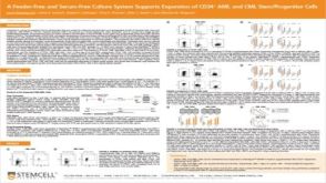 科学海报A Feeder-Free and Serum-Free Culture System Supports Expansion of CD34+ AML and CML Stem/Progenitor Cells


 沪公网安备31010102008431号