Kanai R et al. (JUN 2011)
Clinical cancer research : an official journal of the American Association for Cancer Research 17 11 3686--96
A novel oncolytic herpes simplex virus that synergizes with phosphoinositide 3-kinase/Akt pathway inhibitors to target glioblastoma stem cells.
PURPOSE: To develop a new oncolytic herpes simplex virus (oHSV) for glioblastoma (GBM) therapy that will be effective in glioblastoma stem cells (GSC),an important and untargeted component of GBM. One approach to enhance oHSV efficacy is by combination with other therapeutic modalities. EXPERIMENTAL DESIGN: MG18L,containing a U(S)3 deletion and an inactivating LacZ insertion in U(L)39,was constructed for the treatment of brain tumors. Safety was evaluated after intracerebral injection in HSV-susceptible mice. The efficacy of MG18L in human GSCs and glioma cell lines in vitro was compared with other oHSVs,alone or in combination with phosphoinositide-3-kinase (PI3K)/Akt inhibitors (LY294002,triciribine,GDC-0941,and BEZ235). Cytotoxic interactions between MG18L and PI3K/Akt inhibitors were determined using Chou-Talalay analysis. In vivo efficacy studies were conducted using a clinically relevant mouse model of GSC-derived GBM. RESULTS: MG18L was severely neuroattenuated in mice,replicated well in GSCs,and had anti-GBM activity in vivo. PI3K/Akt inhibitors displayed significant but variable antiproliferative activities in GSCs,whereas their combination with MG18L synergized in killing GSCs and glioma cell lines,but not human astrocytes,through enhanced induction of apoptosis. Importantly,synergy was independent of inhibitor sensitivity. In vivo,the combination of MG18L and LY294002 significantly prolonged survival of mice,as compared with either agent alone,achieving 50% long-term survival in GBM-bearing mice. CONCLUSIONS: This study establishes a novel therapeutic strategy: oHSV manipulation of critical oncogenic pathways to sensitize cancer cells to molecularly targeted drugs. MG18L is a promising agent for the treatment of GBM,being especially effective when combined with PI3K/Akt pathway-targeted agents.
View Publication
产品号#:
05707
产品名:
NeuroCult™化学解离试剂盒(小鼠)
Pease JC et al. (JUL 2012)
Biology open 1 7 622--8
Spontaneous spheroid budding from monolayers: a potential contribution to ovarian cancer dissemination.
Ovarian cancer is the most lethal gynaecologic cancer,in large part because of its early dissemination and rapid development of chemotherapy resistance. Spheroids are clusters of tumor cells found in the peritoneal fluid of patients that are thought to promote this dissemination. Current models suggest that spheroids form by aggregation of single tumor cells shed from the primary tumor. Here,we demonstrate that spheroids can also form by budding directly as adherent clusters from a monolayer. Formation of budded spheroids correlated with expression of vimentin and lack of cortical E-cadherin. We also found that compared to cells grown in monolayers,cells grown as spheroids acquired progressive resistance to the chemotherapy drugs Paclitaxel and Cisplatin. This resistance could be completely reversed by dissociating the spheroids. Our observations highlight a previously unappreciated mode of spheroid formation that might have implications for tumor dissemination and chemotherapy resistance in patients,and suggest that this resistance might be reversed by spheroid dissociation.
View Publication
产品号#:
05620
产品名:
MammoCult™ 人源培养基套装
Dalley AJ et al. (JAN 2013)
Journal of oral pathology & medicine : official publication of the International Association of Oral Pathologists and the American Academy of Oral Pathology 42 1 37--46
Organotypic culture of normal, dysplastic and squamous cell carcinoma-derived oral cell lines reveals loss of spatial regulation of CD44 and p75 NTR in malignancy.
Oral squamous cell carcinomas (OSCC) often arise from dysplastic lesions. The role of cancer stem cells in tumour initiation is widely accepted,yet the potential existence of pre-cancerous stem cells in dysplastic tissue has received little attention. Cell lines from oral diseases ranging in severity from dysplasia to malignancy provide opportunity to investigate the involvement of stem cells in malignant progression from dysplasia. Stem cells are functionally defined by their ability to generate hierarchical tissue structures in consortium with spatial regulation. Organotypic cultures readily display tissue hierarchy in vitro; hence,in this study,we compared hierarchical expression of stem cell-associated markers in dermis-based organotypic cultures of oral epithelial cells from normal tissue (OKF6-TERT2),mild dysplasia (DOK),severe dysplasia (POE-9n) and OSCC (PE/CA P J15). Expression of CD44,p75(NTR),CD24 and ALDH was studied in monolayers by flow cytometry and in organotypic cultures by immunohistochemistry. Spatial regulation of CD44 and p75(NTR) was evident for organotypic cultures of normal (OKF6-TERT2) and dysplasia (DOK and POE-9n) but was lacking for OSCC (PE/CA PJ15)-derived cells. Spatial regulation of CD24 was not evident. All monolayer cultures exhibited CD44,p75(NTR),CD24 antigens and ALDH activity (ALDEFLUOR(®) assay),with a trend towards loss of population heterogeneity that mirrored disease severity. In monolayer,increased FOXA1 and decreased FOXA2 expression correlated with disease severity,but OCT3/4,Sox2 and NANOG did not. We conclude that dermis-based organotypic cultures give opportunity to investigate the mechanisms that underlie loss of spatial regulation of stem cell markers seen with OSCC-derived cells.
View Publication
产品号#:
01700
01705
01702
产品名:
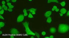
ALDEFLUOR™ 试剂盒
ALDEFLUOR™ DEAB试剂, 1.5 mM, 1 mL
ALDEFLUOR™检测缓冲液
Azari H et al. (JAN 2011)
Journal of visualized experiments : JoVE 56 e3633
Isolation and expansion of human glioblastoma multiforme tumor cells using the neurosphere assay.
Stem-like cells have been isolated in tumors such as breast,lung,colon,prostate and brain. A critical issue in all these tumors,especially in glioblastoma mutliforme (GBM),is to identify and isolate tumor initiating cell population(s) to investigate their role in tumor formation,progression,and recurrence. Understanding tumor initiating cell populations will provide clues to finding effective therapeutic approaches for these tumors. The neurosphere assay (NSA) due to its simplicity and reproducibility has been used as the method of choice for isolation and propagation of many of this tumor cells. This protocol demonstrates the neurosphere culture method to isolate and expand stem-like cells in surgically resected human GBM tumor tissue. The procedures include an initial chemical digestion and mechanical dissociation of tumor tissue,and subsequently plating the resulting single cell suspension in NSA culture. After 7-10 days,primary neurospheres of 150-200 μm in diameter can be observed and are ready for further passaging and expansion.
View Publication
产品号#:
05751
05752
产品名:
NeuroCult™ NS-A 扩增试剂盒(人)
NeuroCult™ NS-A 分化试剂盒(人)
Elling C et al. (MAR 2011)
Blood 117 10 2935--43
Novel imatinib-sensitive PDGFRA-activating point mutations in hypereosinophilic syndrome induce growth factor independence and leukemia-like disease.
The FIP1L1-PDGFRA fusion is seen in a fraction of cases with a presumptive diagnosis of hypereosinophilic syndrome (HES). However,because most HES patients lack FIP1L1-PDGFRA,we studied whether they harbor activating mutations of the PDGFRA gene. Sequencing of 87 FIP1L1-PDGFRA-negative HES patients revealed several novel PDGFRA point mutations (R481G,L507P,I562M,H570R,H650Q,N659S,L705P,R748G,and Y849S). When cloned into 32D cells,N659S and Y849S and-on selection for high expressors-also H650Q and R748G mutants induced growth factor-independent proliferation,clonogenic growth,and constitutive phosphorylation of PDGFRA and Stat5. Imatinib antagonized Stat5 phosphorylation. Mutations involving positions 659 and 849 had been shown previously to possess transforming potential in gastrointestinal stromal tumors. Because H650Q and R748G mutants possessed only weak transforming activity,we injected 32D cells harboring these mutants or FIP1L1-PDGFRA into mice and found that they induced a leukemia-like disease. Oral imatinib treatment significantly decreased leukemic growth in vivo and prolonged survival. In conclusion,our data provide evidence that imatinib-sensitive PDGFRA point mutations play an important role in the pathogenesis of HES and we propose that more research should be performed to further define the frequency and treatment response of PDGFRA mutations in FIP1L1-PDGFRA-negative HES patients.
View Publication
产品号#:
03231
产品名:
MethoCult™ M3231
Penumatsa K et al. (JAN 2010)
Journal of ovarian research 3 28
Differential expression of aldehyde dehydrogenase 1a1 (ALDH1) in normal ovary and serous ovarian tumors.
BACKGROUND: We showed there are specific ALDH1 autoantibodies in ovarian autoimmune disease and ovarian cancer,suggesting a role for ALDH1 in ovarian pathology. However,there is little information on the ovarian expression of ALDH1. Therefore,we compared ALDH1 expression in normal ovary and benign and malignant ovarian tumors to determine if ALDH1 expression is altered in ovarian cancer. Since there is also recent interest in ALDH1 as a cancer stem cell (CSC) marker,we assessed co-expression of ALDH1 with CSC markers in order to determine if ALDH1 is a potential CSC marker in ovarian cancer. METHODS: mRNA and protein expression were compared in normal human ovary and serous ovarian tumors using quantitative Reverse-Transcriptase PCR,Western blot (WB) and semi-quantitative immunohistochemistry (IHC). ALDH1 enzyme activity was confirmed in primary ovarian cells by flow cytometry (FC) using ALDEFLUOR assay. RESULTS: ALDH1 mRNA expression was significantly reduced (p textless 0.01; n = 5) in malignant tumors compared to normal ovaries and benign tumors. The proportion of ALDH1+ cells was significantly lower in malignant tumors (17.1 ± 7.61%; n = 5) compared to normal ovaries (37.4 ± 5.4%; p textless 0.01; n = 5) and benign tumors (31.03 ± 6.68%; p textless 0.05; n = 5). ALDH1+ cells occurred in the stroma and surface epithelium in normal ovary and benign tumors,although surface epithelial expression varied more in benign tumors. Localization of ALDH1 was heterogeneous in malignant tumor cells and little ALDH1 expression occurred in poorly differentiated malignant tumors. In benign tumors the distribution of ALDH1 had features of both normal ovary and malignant tumors. ALDH1 protein expression assessed by IHC,WB and FC was positively correlated (p textless 0.01). ALDH1 did not appear to be co-expressed with the CSC markers CD44,CD117 and CD133 by IHC. CONCLUSIONS: Total ALDH1 expression is significantly reduced in malignant ovarian tumors while it is relatively unchanged in benign tumors compared to normal ovary. Thus,ALDH1 expression in the ovary does not appear to be similar to breast,lung or colon cancer suggesting possible functional differences in these cancers. SIGNIFICANCE: These observations suggest that reduced ALDH1 expression is associated with malignant transformation in ovarian cancer and provides a basis for further study of the mechanism of ALDH1 in this process.
View Publication
产品号#:
01700
01705
07900
01702
100-0762
产品名:
ALDEFLUOR™ 试剂盒
ALDEFLUOR™ DEAB试剂, 1.5 mM, 1 mL
DNase I 溶液(1 mg/mL)
ALDEFLUOR™检测缓冲液
DNase I溶液(1mg /mL)
Denè et al. (SEP 2013)
Clinical chemistry 59 9 1384--92
Capture of viable circulating tumor cells in the liver of colorectal cancer patients.
BACKGROUND The incidence and number of circulating tumor cells (CTCs) in the peripheral blood of colorectal cancer patients are lower than in other cancer types,which may point to a particular biology of colorectal cancer affecting CTC detection. METHODS We detected CTCs in the peripheral and mesenteric blood of colorectal cancer patients by use of 2 independent technologies on the basis of different biological properties of colon cancer cells. Seventy-five patients diagnosed with localized (M0,n = 60) and metastatic (M1,n = 15) colorectal cancer were included. Peripheral and mesenteric blood samples were collected before tumor resection. We performed CTC enumeration with an EpCAM-independent enrichment method followed by the Epispot assay that detected only viable CK19-releasing CTCs. In parallel,we used the FDA-cleared EpCAM-dependent CellSearch® as the reference method. RESULTS The enumeration of CK19-releasing cells by the CK19-Epispot assay revealed viable CTCs in 27 of 41 (65.9%) and 41 of 74 (55.4%) (P = 0.04) patients in mesenteric and peripheral blood,respectively,whereas CellSearch detected CTCs in 19 of 34 (55.9%) and 20 of 69 (29.0%) (P = 0.0046) patients. In mesenteric blood,medians of 4 (range 0-247) and 2.7 CTCs (range 0-286) were found with Epispot and CellSearch (P = 0.2),respectively,whereas in peripheral blood,Epispot and CellSearch detected a median of 1.2 (range 0-92) and 0 CTCs (range 0-147) (P = 0.002). CONCLUSIONS A considerable portion of viable CTCs detectable by the Epispot assay are trapped in the liver as the first filter organ in CRC patients.
View Publication

 EasySep™小鼠TIL(CD45)正选试剂盒


 沪公网安备31010102008431号