Li X et al. (AUG 2012)
Journal of thoracic oncology : official publication of the International Association for the Study of Lung Cancer 7 8 1235--45
Aldehyde dehydrogenase 1A1 possesses stem-like properties and predicts lung cancer patient outcome.
INTRODUCTION: Lung cancer contains a small population of cancer stem cells that contribute to its initiation and progression. We investigated the biological function and clinical significance of aldehyde dehydrogenase 1A1 (ALDH1A1) in non-small-cell lung carcinoma (NSCLC). METHODS: ALDH1A1 assay or small interfering RNA transfection was employed to isolate ALDH1A1+ cells or knock down ALDH1A1 expression in H2087 cells,respectively. Biological functions of ALDH1A1+ and ALDH1A1 silenced cells were investigated using in vitro and in vivo methods. ALDH1A1 expression was analyzed using immunohistochemistry on tissue microarrays with 179 lung cancer tissues and 26 normal lung tissues. RESULTS: The abilities of clone formation,proliferation,cell growth,and migration were increased in ALDH1A1+ and ALDH1A1 silenced cells. ALDH1A1+ lung cancer cells initiated tumors that resembled the histopathologic characteristics and heterogeneity of the parental lung cancer cells in mice. The silencing of ALDH1A1 expression in H2087 lung cancer cells inhibited cell proliferation and migration significantly. ALDH1A1 was expressed in 42% of normal lung tissues (11 of 26),with strong expression in the basal cells and globular cells of the normal bronchus and weak expression in the alveolar epithelial cells. Compared with normal lung tissues,45% of NSCLC samples (81 of 179) were read as positive for ALDH1A1. Positive ALDH1A1 expression was correlated with patients' smoking status (p = 0.022),lymph-node metastasis (p = 0.006),clinical stage (p = 0.004),and a decreased overall survival time (p textless 0.001). Positive ALDH1A1 expression in lung cancer tissues was an independent prognostic factor for NSCLC (odds ratio = 5.232,p textless 0.001). CONCLUSION: Elucidating the biological functions of ALDH1A1 could be helpful in studying lung tumorigenesis and for developing new therapeutic approaches.
View Publication
Maestre-Batlle D et al. (FEB 2017)
Scientific reports 7 42214
Novel flow cytometry approach to identify bronchial epithelial cells from healthy human airways.
Sampling various compartments within the lower airways to examine human bronchial epithelial cells (HBEC) is essential for understanding numerous lung diseases. Conventional methods to identify HBEC in bronchoalveolar lavage (BAL) and wash (BW) have throughput limitations in terms of efficiency and ensuring adequate cell numbers for quantification. Flow cytometry can provide high-throughput quantification of cell number and function in BAL and BW samples,while requiring low cell numbers. To date,a flow cytometric method to identify HBEC recovered from lower human airway samples is unavailable. In this study we present a flow cytometric method identifying HBEC as CD45 negative,EpCAM/pan-cytokeratin (pan-CK) double-positive population after excluding debris,doublets and dead cells from the analysis. For validation,the HBEC panel was applied to primary HBEC resulting in 98.6% of live cells. In healthy volunteers,HBEC recovered from BAL (2.3% of live cells),BW (32.5%) and bronchial brushing samples (88.9%) correlated significantly (p = 0.0001) with the manual microscopy counts with an overall Pearson correlation of 0.96 across the three sample types. We therefore have developed,validated,and applied a flow cytometric method that will be useful to interrogate the role of the respiratory epithelium in multiple lung diseases.
View Publication
产品号#:
05001
05021
05022
05008
产品名:
PneumaCult™-ALI 培养基
PneumaCult™-ALI 培养基含12 mm Transwell®插件
PneumaCult™-ALI 培养基含6.5 mm Transwell®插件
PneumaCult™-Ex 培养基
Prince OA et al. (MAR 2018)
Cellular microbiology 20 3 e12810
Modelling persistent Mycoplasma pneumoniae infection of human airway epithelium.
Mycoplasma pneumoniae is a human respiratory tract pathogen causing acute and chronic airway disease states that can include long-term carriage and extrapulmonary spread. The mechanisms of persistence and migration beyond the conducting airways,however,remain poorly understood. We previously described an acute exposure model using normal human bronchial epithelium (NHBE) in air-liquid interface culture,showing that M. pneumoniae gliding motility is essential for initial colonisation and subsequent spread,including localisation to epithelial cell junctions. We extended those observations here,characterizing M. pneumoniae infection of NHBE for up to 4 weeks. Colonisation of the apical surface was followed by pericellular invasion of the basolateral compartment and migration across the underlying transwell membrane. Despite fluctuations in transepithelial electrical resistance and increased NHBE cell desquamation,barrier function remained largely intact. Desquamation was accompanied by epithelial remodelling that included cytoskeletal reorganisation and development of deep furrows in the epithelium. Finally,M. pneumoniae strains S1 and M129 differed with respect to invasion and histopathology,consistent with contrasting virulence in experimentally infected mice. In summary,this study reports pericellular invasion,NHBE cytoskeletal reorganisation,and tissue remodelling with persistent infection in a human airway epithelium model,providing clear insight into the likely route for extrapulmonary spread.
View Publication


 EasySep™小鼠TIL(CD45)正选试剂盒




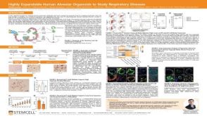
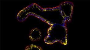
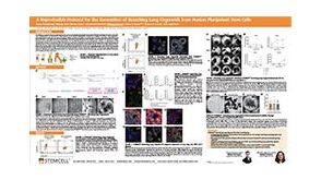 科学海报A Reproducible Protocol for the Generation of Branching Lung Organoids From Human Pluripotent Stem Cells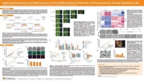


 沪公网安备31010102008431号