Basma H et al. (MAR 2014)
American journal of physiology. Lung cellular and molecular physiology 306 6 L552--65
Reprogramming of COPD lung fibroblasts through formation of induced pluripotent stem cells.
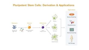
Reprogramming somatic cells to induced pluripotent stem cells (iPSCs) eliminates many epigenetic modifications that characterize differentiated cells. In this study,we tested whether functional differences between chronic obstructive pulmonary disease (COPD) and non-COPD fibroblasts could be reduced utilizing this approach. Primary fibroblasts from non-COPD and COPD patients were reprogrammed to iPSCs. Reprogrammed iPSCs were positive for oct3/4,nanog,and sox2,formed embryoid bodies in vitro,and induced teratomas in nonobese diabetic/severe combined immunodeficient mice. Reprogrammed iPSCs were then differentiated into fibroblasts (non-COPD-i and COPD-i) and were assessed either functionally by chemotaxis and gel contraction or for gene expression by microarrays and compared with their corresponding primary fibroblasts. Primary COPD fibroblasts contracted three-dimensional collagen gels and migrated toward fibronectin less robustly than non-COPD fibroblasts. In contrast,redifferentiated fibroblasts from iPSCs derived from the non-COPD and COPD fibroblasts were similar in response in both functional assays. Microarray analysis identified 1,881 genes that were differentially expressed between primary COPD and non-COPD fibroblasts,with 605 genes differing by more than twofold. After redifferentiation,112 genes were differentially expressed between COPD-i and non-COPD-i with only three genes by more than twofold. Similar findings were observed with microRNA (miRNA) expression: 56 miRNAs were differentially expressed between non-COPD and COPD primary cells; after redifferentiation,only 3 miRNAs were differentially expressed between non-COPD-i and COPD-i fibroblasts. Interestingly,of the 605 genes that were differentially expressed between COPD and non-COPD fibroblasts,293 genes were changed toward control after redifferentiation. In conclusion,functional and epigenetic alterations of COPD fibroblasts can be reprogrammed through formation of iPSCs.
View Publication
产品号#:
05850
05857
05870
05875
85850
85857
85870
85875
产品名:
mTeSR™1
mTeSR™1
Deng M et al. (JAN 2018)
European Journal of Neuroscience 47 2 150--157
Preservation of neuronal functions by exosomes derived from different human neural cell types under ischemic conditions
Stem cell-based therapies have been reported in protecting cerebral infarction-induced neuronal dysfunction and death. However,most studies used rat/mouse neuron as model cell when treated with stem cell or exosomes. Whether these findings can be translated from rodent to humans has been in doubt. Here,we used human embryonic stem cell-derived neurons to detect the protective potential of exosomes against ischemia. Neurons were treated with in vitro oxygen-glucose deprivation (OGD) for 1 h. For treatment group,different exosomes were derived from neuron,embryonic stem cell,neural progenitor cell and astrocyte differentiated from H9 human embryonic stem cell and added to culture medium 30 min after OGD (100 μg/mL). Western blotting was performed 12 h after OGD,while cell counting and electrophysiological recording were performed 48 h after OGD. We found that these exosomes attenuated OGD-induced neuronal death,Mammalian target of rapamycin (mTOR),pro-inflammatory and apoptotic signaling pathway changes,as well as basal spontaneous synaptic transmission inhibition in varying degrees. The results implicate the protective effect of exosomes on OGD-induced neuronal death and dysfunction in human embryonic stem cell-derived neurons,potentially through their modulation on mTOR,pro-inflammatory and apoptotic signaling pathways.
View Publication
产品号#:
05711
05790
05792
05793
05794
05795
100-1281
产品名:
NeuroCult™ SM1 神经添加物
BrainPhys™神经元培养基
BrainPhys™神经元培养基和SM1试剂盒
BrainPhys™ 神经元培养基N2-A和SM1试剂盒
BrainPhys™原代神经元试剂盒
BrainPhys™ hPSC 神经元试剂盒
NeuroCult™ SM1 神经添加物
Lippmann ES et al. (APR 2014)
Stem Cells 32 4 1032--1042
Defined human pluripotent stem cell culture enables highly efficient neuroepithelium derivation without small molecule inhibitors.
The embryonic neuroepithelium gives rise to the entire central nervous system in vivo,making it an important tissue for developmental studies and a prospective cell source for regenerative applications. Current protocols for deriving homogenous neuroepithelial cultures from human pluripotent stem cells (hPSCs) consist of either embryoid body-mediated neuralization followed by a manual isolation step or adherent differentiation using small molecule inhibitors. Here,we report that hPSCs maintained under chemically defined,feeder-independent,and xeno-free conditions can be directly differentiated into pure neuroepithelial cultures ([mt]90% Pax6(+)/N-cadherin(+) with widespread rosette formation) within 6 days under adherent conditions,without small molecule inhibitors,and using only minimalistic medium consisting of Dulbecco's modified Eagle's medium/F-12,sodium bicarbonate,selenium,ascorbic acid,transferrin,and insulin (i.e.,E6 medium). Furthermore,we provide evidence that the defined culture conditions enable this high level of neural conversion in contrast to hPSCs maintained on mouse embryonic fibroblasts (MEFs). In addition,hPSCs previously maintained on MEFs could be rapidly converted to a neural compliant state upon transfer to these defined conditions while still maintaining their ability to generate all three germ layers. Overall,this fully defined and scalable protocol should be broadly useful for generating therapeutic neural cells for regenerative applications.
View Publication

 EasySep™小鼠TIL(CD45)正选试剂盒


 27:19
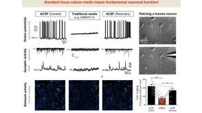
线上讲座BrainPhys™ Medium Supports the Physiological Activity of Neuronal Tissue in vitro发布日期: 07/22/2016
27:19
线上讲座BrainPhys™ Medium Supports the Physiological Activity of Neuronal Tissue in vitro发布日期: 07/22/2016

 沪公网安备31010102008431号