Werner A et al. (SEP 2015)
Nature 525 7570 523--527
Cell-fate determination by ubiquitin-dependent regulation of translation
Metazoan development depends on the accurate execution of differentiation programs that allow pluripotent stem cells to adopt specific fates. Differentiation requires changes to chromatin architecture and transcriptional networks,yet whether other regulatory events support cell-fate determination is less well understood. Here we identify the ubiquitin ligase CUL3 in complex with its vertebrate-specific substrate adaptor KBTBD8 (CUL3(KBTBD8)) as an essential regulator of human and Xenopus tropicalis neural crest specification. CUL3(KBTBD8) monoubiquitylates NOLC1 and its paralogue TCOF1,the mutation of which underlies the neurocristopathy Treacher Collins syndrome. Ubiquitylation drives formation of a TCOF1-NOLC1 platform that connects RNA polymerase I with ribosome modification enzymes and remodels the translational program of differentiating cells in favour of neural crest specification. We conclude that ubiquitin-dependent regulation of translation is an important feature of cell-fate determination.
View Publication
产品号#:
05872
05873
07920
07922
05210
05215
34811
34815
34850
34821
34825
34860
05835
05839
100-0483
100-0484
产品名:
ACCUTASE™
ACCUTASE™
AggreWell™ 800 24孔板,1个
AggreWell™ 800 24孔板,5个
AggreWell™ 800 24孔板启动套装
AggreWell™ 800 6孔板,1个
AggreWell™ 800 6孔板,5个
AggreWell™ 800 6孔板启动套装
STEMdiff™ 神经诱导培养基
STEMdiff™ 神经诱导培养基
Hausser Scientificᵀᴹ 明线血球计数板
ReLeSR™
Yamazaki K et al. (DEC 2016)
Journal of Biomolecular Screening 21 10 1054--1064
Functional Comparison of Neuronal Cells Differentiated from Human Induced Pluripotent Stem CellDerived Neural Stem Cells under Different Oxygen and Medium Conditions
Because neurons are difficult to obtain from humans,generating functional neurons from human induced pluripotent stem cells (hiPSCs) is important for establishing physiological or disease-relevant screening systems for drug discovery. To examine the culture conditions leading to efficient differentiation of functional neural cells,we investigated the effects of oxygen stress (2% or 20% O2) and differentiation medium (DMEM/F12:Neurobasal-based [DN] or commercial [PhoenixSongs Biologicals; PS]) on the expression of genes related to neural differentiation,glutamate receptor function,and the formation of networks of neurons differentiated from hiPSCs (201B7) via long-term self-renewing neuroepithelial-like stem (lt-NES) cells. Expression of genes related to neural differentiation occurred more quickly in PS and/or 2% O2 than in DN and/or 20% O2,resulting in high responsiveness of neural cells to glutamate,N-methyl-d-aspartate (NMDA),α-amino-3-hydroxy-5-methyl-4-isoxazolepropionate (AMPA),and (S)-3,5-d...
View Publication
产品号#:
05832
产品名:
STEMdiff™ 神经花环选择试剂
Lippmann ES et al. (APR 2014)
Stem Cells 32 4 1032--1042
Defined human pluripotent stem cell culture enables highly efficient neuroepithelium derivation without small molecule inhibitors.
The embryonic neuroepithelium gives rise to the entire central nervous system in vivo,making it an important tissue for developmental studies and a prospective cell source for regenerative applications. Current protocols for deriving homogenous neuroepithelial cultures from human pluripotent stem cells (hPSCs) consist of either embryoid body-mediated neuralization followed by a manual isolation step or adherent differentiation using small molecule inhibitors. Here,we report that hPSCs maintained under chemically defined,feeder-independent,and xeno-free conditions can be directly differentiated into pure neuroepithelial cultures ([mt]90% Pax6(+)/N-cadherin(+) with widespread rosette formation) within 6 days under adherent conditions,without small molecule inhibitors,and using only minimalistic medium consisting of Dulbecco's modified Eagle's medium/F-12,sodium bicarbonate,selenium,ascorbic acid,transferrin,and insulin (i.e.,E6 medium). Furthermore,we provide evidence that the defined culture conditions enable this high level of neural conversion in contrast to hPSCs maintained on mouse embryonic fibroblasts (MEFs). In addition,hPSCs previously maintained on MEFs could be rapidly converted to a neural compliant state upon transfer to these defined conditions while still maintaining their ability to generate all three germ layers. Overall,this fully defined and scalable protocol should be broadly useful for generating therapeutic neural cells for regenerative applications.
View Publication
A homozygous loss-of-function CAMK2A mutation causes growth delay, frequent seizures and severe intellectual disability.
Calcium/calmodulin-dependent protein kinase II (CAMK2) plays fundamental roles in synaptic plasticity that underlies learning and memory. Here,we describe a new recessive neurodevelopmental syndrome with global developmental delay,seizures and intellectual disability. Using linkage analysis and exome sequencing,we found that this disease maps to chromosome 5q31.1-q34 and is caused by a biallelic germline mutation in CAMK2A. The missense mutation,p.His477Tyr is located in the CAMK2A association domain that is critical for its function and localization. Biochemically,the p.His477Tyr mutant is defective in self-oligomerization and unable to assemble into the multimeric holoenzyme.In vivo,CAMK2AH477Y failed to rescue neuronal defects in C. elegans lacking unc-43,the ortholog of human CAMK2A. In vitro,neurons derived from patient iPSCs displayed profound synaptic defects. Together,our data demonstrate that a recessive germline mutation in CAMK2A leads to neurodevelopmental defects in humans and suggest that dysfunctional CAMK2 paralogs may contribute to other neurological disorders.
View Publication
产品号#:
05790
05792
05793
05794
05795
85850
85857
85870
85875
产品名:
BrainPhys™神经元培养基
BrainPhys™神经元培养基和SM1试剂盒
BrainPhys™ 神经元培养基N2-A和SM1试剂盒
BrainPhys™原代神经元试剂盒
BrainPhys™ hPSC 神经元试剂盒
mTeSR™1
mTeSR™1
Xia G et al. (JUN 2015)
Stem cells (Dayton,Ohio) 33 6 1829--38
Genome modification leads to phenotype reversal in human myotonic dystrophy type 1 induced pluripotent stem cell-derived neural stem cells.
Myotonic dystrophy type 1 (DM1) is caused by expanded CTG repeats in the 3'-untranslated region (3' UTR) of the DMPK gene. Correcting the mutation in DM1 stem cells would be an important step toward autologous stem cell therapy. The objective of this study is to demonstrate in vitro genome editing to prevent production of toxic mutant transcripts and reverse phenotypes in DM1 stem cells. Genome editing was performed in DM1 neural stem cells (NSCs) derived from human DM1 induced pluripotent stem (iPS) cells. An editing cassette containing SV40/bGH polyA signals was integrated upstream of the CTG repeats by TALEN-mediated homologous recombination (HR). The expression of mutant CUG repeats transcript was monitored by nuclear RNA foci,the molecular hallmarks of DM1,using RNA fluorescence in situ hybridization. Alternative splicing of microtubule-associated protein tau (MAPT) and muscleblind-like (MBNL) proteins were analyzed to further monitor the phenotype reversal after genome modification. The cassette was successfully inserted into DMPK intron 9 and this genomic modification led to complete disappearance of nuclear RNA foci. MAPT and MBNL 1,2 aberrant splicing in DM1 NSCs were reversed to normal pattern in genome-modified NSCs. Genome modification by integration of exogenous polyA signals upstream of the DMPK CTG repeat expansion prevents the production of toxic RNA and leads to phenotype reversal in human DM1 iPS-cells derived stem cells. Our data provide proof-of-principle evidence that genome modification may be used to generate genetically modified progenitor cells as a first step toward autologous cell transfer therapy for DM1.
View Publication
产品号#:
05833
05835
05839
产品名:
STEMdiff™神经前体细胞培养基
STEMdiff™ 神经诱导培养基
STEMdiff™ 神经诱导培养基
M. van den Hurk et al. ( 2018)
Frontiers in Molecular Neuroscience
Patch-Seq Protocol to Analyze the Electrophysiology, Morphology and Transcriptome of Whole Single Neurons Derived From Human Pluripotent Stem Cells
The human brain is composed of a complex assembly of about 171 billion heterogeneous cellular units (86 billion neurons and 85 billion non-neuronal glia cells). A comprehensive description of brain cells is necessary to understand the nervous system in health and disease. Recently,advances in genomics have permitted the accurate analysis of the full transcriptome of single cells (scRNA-seq). We have built upon such technical progress to combine scRNA-seq with patch-clamping electrophysiological recording and morphological analysis of single human neurons in vitro. This new powerful method,referred to as Patch-seq,enables a thorough,multimodal profiling of neurons and permits us to expose the links between functional properties,morphology,and gene expression. Here,we present a detailed Patch-seq protocol for isolating single neurons from in vitro neuronal cultures. We have validated the Patch-seq whole-transcriptome profiling method with human neurons generated from embryonic and induced pluripotent stem cells (ESCs/iPSCs) derived from healthy subjects,but the procedure may be applied to any kind of cell type in vitro. Patch-seq may be used on neurons in vitro to profile cell types and states in depth to unravel the human molecular basis of neuronal diversity and investigate the cellular mechanisms underlying brain disorders.
View Publication
产品号#:
05711
07152
07920
07922
05790
05792
05793
05794
05795
100-1281
产品名:
NeuroCult™ SM1 神经添加物
N2 添加物-A
ACCUTASE™
ACCUTASE™
BrainPhys™神经元培养基
BrainPhys™神经元培养基和SM1试剂盒
BrainPhys™ 神经元培养基N2-A和SM1试剂盒
BrainPhys™原代神经元试剂盒
BrainPhys™ hPSC 神经元试剂盒
NeuroCult™ SM1 神经添加物
C. L. Moreno et al. ( 2018)
Molecular neurodegeneration 13 1 33
BACKGROUND Type 2 diabetes (T2D) is a recognized risk factor for the development of cognitive impairment (CI) and/or dementia,although the exact nature of the molecular pathology of T2D-associated CI remains obscure. One link between T2D and CI might involve decreased insulin signaling in brain and/or neurons in either animal or postmortem human brains as has been reported as a feature of Alzheimer's disease (AD). Here we asked if neuronal insulin resistance is a cell autonomous phenomenon in a familial form of AD. METHODS We have applied a newly developed protocol for deriving human basal forebrain cholinergic neurons (BFCN) from skin fibroblasts via induced pluripotent stem cell (iPSC) technology. We generated wildtype and familial AD mutant PSEN2 N141I (presenilin 2) BFCNs and assessed if insulin signaling,insulin regulation of the major AD proteins Abeta$ and/or tau,and/or calcium fluxes is altered by the PSEN2 N141I mutation. RESULTS We report herein that wildtype,PSEN2 N141I and CRISPR/Cas9-corrected iPSC-derived BFCNs (and their precursors) show indistinguishable insulin signaling profiles as determined by the phosphorylation of canonical insulin signaling pathway molecules. Chronic insulin treatment of BFCNs of all genotypes led to a reduction in the Abeta$42/40 ratio. Unexpectedly,we found a CRISPR/Cas9-correctable effect of PSEN2 N141I on calcium flux,which could be prevented by chronic exposure of BFCNs to insulin. CONCLUSIONS Our studies indicate that the familial AD mutation PSEN2 N141I does not induce neuronal insulin resistance in a cell autonomous fashion. The ability of insulin to correct calcium fluxes and to lower Abeta$42/40 ratio suggests that insulin acts to oppose an AD-pathophysiology. Hence,our results are consistent with a potential physiological role for insulin as a mediator of resilience by counteracting specific metabolic and molecular features of AD.
View Publication
Deng M et al. (JAN 2018)
European Journal of Neuroscience 47 2 150--157
Preservation of neuronal functions by exosomes derived from different human neural cell types under ischemic conditions
Stem cell-based therapies have been reported in protecting cerebral infarction-induced neuronal dysfunction and death. However,most studies used rat/mouse neuron as model cell when treated with stem cell or exosomes. Whether these findings can be translated from rodent to humans has been in doubt. Here,we used human embryonic stem cell-derived neurons to detect the protective potential of exosomes against ischemia. Neurons were treated with in vitro oxygen-glucose deprivation (OGD) for 1 h. For treatment group,different exosomes were derived from neuron,embryonic stem cell,neural progenitor cell and astrocyte differentiated from H9 human embryonic stem cell and added to culture medium 30 min after OGD (100 μg/mL). Western blotting was performed 12 h after OGD,while cell counting and electrophysiological recording were performed 48 h after OGD. We found that these exosomes attenuated OGD-induced neuronal death,Mammalian target of rapamycin (mTOR),pro-inflammatory and apoptotic signaling pathway changes,as well as basal spontaneous synaptic transmission inhibition in varying degrees. The results implicate the protective effect of exosomes on OGD-induced neuronal death and dysfunction in human embryonic stem cell-derived neurons,potentially through their modulation on mTOR,pro-inflammatory and apoptotic signaling pathways.
View Publication

 EasySep™小鼠TIL(CD45)正选试剂盒


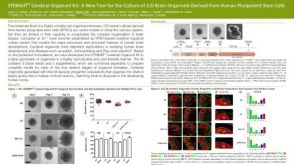 科学海报STEMdiff™ Cerebral Organoid Kit: A New Tool for the Culture of 3D Brain Organoids Derived from hPSCs


 沪公网安备31010102008431号