Y. Kim et al. (May 2020)
FASEB Journal 34 6965-6983
Microtubule-associated protein 2 mediates induction of long-term potentiation in hippocampal neurons
Microtubule-associated protein (MAP) 2 has been perceived as a static cytoskeletal protein enriched in neuronal dendritic shafts. Emerging evidence indicates dynamic functions for various MAPs in activity-dependent synaptic plasticity. However,it is unclear how MAP2 is associated with synaptic plasticity mechanisms. Here,we demonstrate that specific silencing of high-molecular-weight MAP2 in vivo abolished induction of long-term potentiation (LTP) in the Schaffer collateral pathway of CA1 pyramidal neurons and in vitro blocked LTP-induced surface delivery of AMPA receptors and spine enlargement. In mature hippocampal neurons,we observed rapid translocation of a subpopulation of MAP2,present in dendritic shafts,to spines following LTP stimulation. Time-lapse confocal imaging showed that spine translocation of MAP2 was coupled with LTP-induced spine enlargement. Consistently,immunogold electron microscopy revealed that LTP stimulation of the Schaffer collateral pathway promoted MAP2 labeling in spine heads of CA1 neurons. This translocation depended on NMDA receptor activation and Ras-MAPK signaling. Furthermore,LTP stimulation led to an increase in surface-expressed AMPA receptors specifically in the neurons with MAP2 spine translocation. Altogether,this study indicates a novel role for MAP2 in LTP mechanisms and suggests that MAP2 participates in activity-dependent synaptic plasticity in mature hippocampal networks.
View Publication
Katori S et al. (JUL 2009)
The Journal of neuroscience : the official journal of the Society for Neuroscience 29 29 9137--47
Protocadherin-alpha family is required for serotonergic projections to appropriately innervate target brain areas.
Serotonergic axons from the raphe nuclei in the brainstem project to every region of the brain,where they make connections through their extensive terminal arborizations. This serotonergic innervation contributes to various normal behaviors and psychiatric disorders. The protocadherin-alpha (Pcdha) family of clustered protocadherins consists of 14 cadherin-related molecules generated from a single gene cluster. We found that the Pcdhas were strongly expressed in the serotonergic neurons. To elucidate their roles,we examined serotonergic fibers in a mouse mutant (Pcdha(Delta CR/Delta CR)) lacking the Pcdha cytoplasmic region-encoding exons,which are common to the gene cluster. In the first week after birth,the distribution pattern of serotonergic fibers in Pcdha(Delta CR/Delta CR) mice was similar to wild-type,but by 3 weeks of age,when the serotonergic axonal termini complete their arborizations,the distribution of the projections was abnormal. In some target regions,notably the globus pallidus and substantia nigra,the normally even distribution of serotonin axonal terminals was,in the mutants,dense at the periphery of each region,but sparse in the center. In the stratum lacunosum-molecular of the hippocampus,the mutants showed denser serotonergic innervation than in wild-type,and in the dentate gyrus of the hippocampus and the caudate-putamen,the innervation was sparser. Together,the abnormalities suggested that Pcdha proteins are important in the late-stage maturation of serotonergic projections. Further examination of alternatively spliced exons encoding the cytoplasmic tail showed that the A-type (but not the B-type) cytoplasmic tail was essential for the normal development of serotonergic projections.
View Publication
产品号#:
03800
03801
03802
03803
03804
03805
03806
产品名:
ClonaCell™-HY杂交瘤试剂盒
ClonaCell™-HY培养基A
ClonaCell™-HY 培养基 B
ClonaCell™-HY 培养基 C
ClonaCell™-HY 培养基 D
ClonaCell™-HY 培养基 E
ClonaCell™-HY PEG
Jackson TC et al. (FEB 2018)
Experimental Neurology 300 232--246
BrainPhys increases neurofilament levels in CNS cultures, and facilitates investigation of axonal damage after a mechanical stretch-injury in vitro
Neurobasal®/B27 is a gold standard culture media used to study primary neurons in vitro. An alternative media (BrainPhys®/SM1) was recently developed which robustly enhances neuronal activity vs. Neurobasal® or DMEM. To the best of our knowledge BrainPhys® has not been explored in the setting of neuronal injury. Here we characterized the utility of BrainPhys® in a model of in vitro mechanical-stretch injury. METHODS/RESULTSPrimary rat cortical neurons were maintained in classic Neurobasal®,or sequentially maintained in Neurocult® followed by BrainPhys® (hereafter simply referred to as BrainPhys® maintained neurons?). The levels of axonal markers and proteins involved in neurotransmission were compared on day in vitro 10 (DIV10). BrainPhys® maintained neurons had higher levels of GluN2B,GluR1,Neurofilament light/heavy chain (NF-L & NF-H),and protein phosphatase 2 A (PP2A) vs. neurons in Neurobasal®. Mechanical stretch-injury (50ms/54% biaxial stretch) to BrainPhys® maintained neurons modestly (albeit significantly) increased 24h lactate dehydrogenase (LDH) levels but markedly decreased axonal NF-L levels post-injury vs. uninjured controls or neurons given a milder 38% stretch-injury. Furthermore,two 54% stretch-injuries (in tandem) exacerbated 24h LDH release,increased α-spectrin breakdown products (SBDPs),and decreased Tau levels. Also,BrainPhys® maintained cultures had decreased markers of cell damage 24h after a single 54% stretch-injury vs. neurons in Neurobasal®. Finally,we tested the hypothesis that lentivirus mediated overexpression of the pro-death protein RBM5 exacerbates neuronal and/or axonal injury in primary CNS cultures. RBM5 overexpression vs. empty-vector controls increased 24h LDH release,and SBDP levels,after a single 54% stretch-injury but did not affect NF-L levels or Tau. CONCLUSIONBrainPhys® is a promising new reagent which facilities the investigation of molecular targets involved in axonal and/or neuronal injury in vitro.
View Publication
产品号#:
05790
05792
05793
05794
05795
产品名:
BrainPhys™神经元培养基
BrainPhys™神经元培养基和SM1试剂盒
BrainPhys™ 神经元培养基N2-A和SM1试剂盒
BrainPhys™原代神经元试剂盒
BrainPhys™ hPSC 神经元试剂盒
Jessick VJ et al. ( 2013)
International journal of physiology,pathophysiology and pharmacology 5 4 216--27
Investigating the role of the actin regulating complex ARP2/3 in rapid ischemic tolerance induced neuro-protection.
Neuronal morphology is highly sensitive to ischemia,although some re-organization may promote neuroprotection. In this study we investigate the role of actin regulating proteins (ARP2,ARP3 and WAVE-1) and their role in rapid ischemic tolerance. Using an established in vitro model of rapid ischemic tolerance,we show that WAVE-1 protein levels are stabilized following brief tolerance inducing ischemia (preconditioning). The stabilization appears to be due to a reduction in the ubiquitination of WAVE-1. Levels of ARP2,ARP3 and N-WASP were not affected by ischemic preconditioning. Immunocytochemical studies show a relocalization of ARP2 and ARP3 proteins in neurons following preconditioning ischemia,as well as a re-organization of actin. Blocking the protein kinase CK2 using emodin blocks ischemic tolerance,and our data suggests CK2 binds to WAVE-1 in neurons. We observe an increase in binding of the ARP2 subunit with WAVE-1. The neuroprotection observed following preconditioning is inhibited when cells are transduced with an N-WASP CA domain that blocks the activation of ARP2/3. Together these data show that ischemia affects actin regulating enzymes,and that the ARP2/3 pathway plays a role in rapid ischemic tolerance induced neuroprotection.
View Publication

 EasySep™小鼠TIL(CD45)正选试剂盒


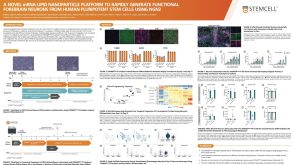 科学海报A Novel mRNA-LNP Platform to Rapidly Generate Functional Forebrain Neurons from hPSCs Using NGN2


 沪公网安备31010102008431号