Yamazaki K et al. (DEC 2016)
Journal of Biomolecular Screening 21 10 1054--1064
Functional Comparison of Neuronal Cells Differentiated from Human Induced Pluripotent Stem CellDerived Neural Stem Cells under Different Oxygen and Medium Conditions
Because neurons are difficult to obtain from humans,generating functional neurons from human induced pluripotent stem cells (hiPSCs) is important for establishing physiological or disease-relevant screening systems for drug discovery. To examine the culture conditions leading to efficient differentiation of functional neural cells,we investigated the effects of oxygen stress (2% or 20% O2) and differentiation medium (DMEM/F12:Neurobasal-based [DN] or commercial [PhoenixSongs Biologicals; PS]) on the expression of genes related to neural differentiation,glutamate receptor function,and the formation of networks of neurons differentiated from hiPSCs (201B7) via long-term self-renewing neuroepithelial-like stem (lt-NES) cells. Expression of genes related to neural differentiation occurred more quickly in PS and/or 2% O2 than in DN and/or 20% O2,resulting in high responsiveness of neural cells to glutamate,N-methyl-d-aspartate (NMDA),α-amino-3-hydroxy-5-methyl-4-isoxazolepropionate (AMPA),and (S)-3,5-d...
View Publication
产品号#:
05832
产品名:
STEMdiff™ 神经花环选择试剂
Deng M et al. (JAN 2018)
European Journal of Neuroscience 47 2 150--157
Preservation of neuronal functions by exosomes derived from different human neural cell types under ischemic conditions
Stem cell-based therapies have been reported in protecting cerebral infarction-induced neuronal dysfunction and death. However,most studies used rat/mouse neuron as model cell when treated with stem cell or exosomes. Whether these findings can be translated from rodent to humans has been in doubt. Here,we used human embryonic stem cell-derived neurons to detect the protective potential of exosomes against ischemia. Neurons were treated with in vitro oxygen-glucose deprivation (OGD) for 1 h. For treatment group,different exosomes were derived from neuron,embryonic stem cell,neural progenitor cell and astrocyte differentiated from H9 human embryonic stem cell and added to culture medium 30 min after OGD (100 μg/mL). Western blotting was performed 12 h after OGD,while cell counting and electrophysiological recording were performed 48 h after OGD. We found that these exosomes attenuated OGD-induced neuronal death,Mammalian target of rapamycin (mTOR),pro-inflammatory and apoptotic signaling pathway changes,as well as basal spontaneous synaptic transmission inhibition in varying degrees. The results implicate the protective effect of exosomes on OGD-induced neuronal death and dysfunction in human embryonic stem cell-derived neurons,potentially through their modulation on mTOR,pro-inflammatory and apoptotic signaling pathways.
View Publication
产品号#:
05711
05790
05792
05793
05794
05795
100-1281
产品名:
NeuroCult™ SM1 神经添加物
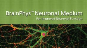
BrainPhys™神经元培养基
BrainPhys™神经元培养基和SM1试剂盒
BrainPhys™ 神经元培养基N2-A和SM1试剂盒
BrainPhys™原代神经元试剂盒
BrainPhys™ hPSC 神经元试剂盒
NeuroCult™ SM1 神经添加物
S. Bell et al. (JUL 2018)
Stem cell reports 11 1 183--196
Disruption of GRIN2B Impairs Differentiation in Human Neurons.
Heterozygous loss-of-function mutations in GRIN2B,a subunit of the NMDA receptor,cause intellectual disability and language impairment. We developed clonal models of GRIN2B deletion and loss-of-function mutations in a region coding for the glutamate binding domain in human cells and generated neurons from a patient harboring a missense mutation in the same domain. Transcriptome analysis revealed extensive increases in genes associated with cell proliferation and decreases in genes associated with neuron differentiation,a result supported by extensive protein analyses. Using electrophysiology and calcium imaging,we demonstrate that NMDA receptors are present on neural progenitor cells and that human mutations in GRIN2B can impair calcium influx and membrane depolarization even in a presumed undifferentiated cell state,highlighting an important role for non-synaptic NMDA receptors. It may be this function,in part,which underlies the neurological disease observed in patients with GRIN2B mutations.
View Publication
产品号#:
05833
05872
05873
05910
07174
05790
05792
05793
05794
05795
85850
85857
85870
85875
100-0483
100-0484
05914
100-0485
100-1077
产品名:
STEMdiff™神经前体细胞培养基
BrainPhys™神经元培养基
BrainPhys™神经元培养基和SM1试剂盒
BrainPhys™ 神经元培养基N2-A和SM1试剂盒
BrainPhys™原代神经元试剂盒
BrainPhys™ hPSC 神经元试剂盒
mTeSR™1
mTeSR™1
Hausser Scientificᵀᴹ 明线血球计数板
ReLeSR™
TeSR™-E7™重编程培养基(2组分)
温和细胞解离试剂
ReLeSR™
P. H. Chia et al. (MAY 2018)
eLife 7
A homozygous loss-of-function CAMK2A mutation causes growth delay, frequent seizures and severe intellectual disability.
Calcium/calmodulin-dependent protein kinase II (CAMK2) plays fundamental roles in synaptic plasticity that underlies learning and memory. Here,we describe a new recessive neurodevelopmental syndrome with global developmental delay,seizures and intellectual disability. Using linkage analysis and exome sequencing,we found that this disease maps to chromosome 5q31.1-q34 and is caused by a biallelic germline mutation in CAMK2A. The missense mutation,p.His477Tyr is located in the CAMK2A association domain that is critical for its function and localization. Biochemically,the p.His477Tyr mutant is defective in self-oligomerization and unable to assemble into the multimeric holoenzyme.In vivo,CAMK2AH477Y failed to rescue neuronal defects in C. elegans lacking unc-43,the ortholog of human CAMK2A. In vitro,neurons derived from patient iPSCs displayed profound synaptic defects. Together,our data demonstrate that a recessive germline mutation in CAMK2A leads to neurodevelopmental defects in humans and suggest that dysfunctional CAMK2 paralogs may contribute to other neurological disorders.
View Publication
产品号#:
05790
05792
05793
05794
05795
85850
85857
85870
85875
产品名:
BrainPhys™神经元培养基
BrainPhys™神经元培养基和SM1试剂盒
BrainPhys™ 神经元培养基N2-A和SM1试剂盒
BrainPhys™原代神经元试剂盒
BrainPhys™ hPSC 神经元试剂盒
mTeSR™1
mTeSR™1
C. L. Moreno et al. ( 2018)
Molecular neurodegeneration 13 1 33
BACKGROUND Type 2 diabetes (T2D) is a recognized risk factor for the development of cognitive impairment (CI) and/or dementia,although the exact nature of the molecular pathology of T2D-associated CI remains obscure. One link between T2D and CI might involve decreased insulin signaling in brain and/or neurons in either animal or postmortem human brains as has been reported as a feature of Alzheimer's disease (AD). Here we asked if neuronal insulin resistance is a cell autonomous phenomenon in a familial form of AD. METHODS We have applied a newly developed protocol for deriving human basal forebrain cholinergic neurons (BFCN) from skin fibroblasts via induced pluripotent stem cell (iPSC) technology. We generated wildtype and familial AD mutant PSEN2 N141I (presenilin 2) BFCNs and assessed if insulin signaling,insulin regulation of the major AD proteins Abeta$ and/or tau,and/or calcium fluxes is altered by the PSEN2 N141I mutation. RESULTS We report herein that wildtype,PSEN2 N141I and CRISPR/Cas9-corrected iPSC-derived BFCNs (and their precursors) show indistinguishable insulin signaling profiles as determined by the phosphorylation of canonical insulin signaling pathway molecules. Chronic insulin treatment of BFCNs of all genotypes led to a reduction in the Abeta$42/40 ratio. Unexpectedly,we found a CRISPR/Cas9-correctable effect of PSEN2 N141I on calcium flux,which could be prevented by chronic exposure of BFCNs to insulin. CONCLUSIONS Our studies indicate that the familial AD mutation PSEN2 N141I does not induce neuronal insulin resistance in a cell autonomous fashion. The ability of insulin to correct calcium fluxes and to lower Abeta$42/40 ratio suggests that insulin acts to oppose an AD-pathophysiology. Hence,our results are consistent with a potential physiological role for insulin as a mediator of resilience by counteracting specific metabolic and molecular features of AD.
View Publication
Lippmann ES et al. (APR 2014)
Stem Cells 32 4 1032--1042
Defined human pluripotent stem cell culture enables highly efficient neuroepithelium derivation without small molecule inhibitors.
The embryonic neuroepithelium gives rise to the entire central nervous system in vivo,making it an important tissue for developmental studies and a prospective cell source for regenerative applications. Current protocols for deriving homogenous neuroepithelial cultures from human pluripotent stem cells (hPSCs) consist of either embryoid body-mediated neuralization followed by a manual isolation step or adherent differentiation using small molecule inhibitors. Here,we report that hPSCs maintained under chemically defined,feeder-independent,and xeno-free conditions can be directly differentiated into pure neuroepithelial cultures ([mt]90% Pax6(+)/N-cadherin(+) with widespread rosette formation) within 6 days under adherent conditions,without small molecule inhibitors,and using only minimalistic medium consisting of Dulbecco's modified Eagle's medium/F-12,sodium bicarbonate,selenium,ascorbic acid,transferrin,and insulin (i.e.,E6 medium). Furthermore,we provide evidence that the defined culture conditions enable this high level of neural conversion in contrast to hPSCs maintained on mouse embryonic fibroblasts (MEFs). In addition,hPSCs previously maintained on MEFs could be rapidly converted to a neural compliant state upon transfer to these defined conditions while still maintaining their ability to generate all three germ layers. Overall,this fully defined and scalable protocol should be broadly useful for generating therapeutic neural cells for regenerative applications.
View Publication
产品号#:
05850
05857
05870
05875
85850
85857
85870
85875
产品名:
mTeSR™1
mTeSR™1
Basma H et al. (MAR 2014)
American journal of physiology. Lung cellular and molecular physiology 306 6 L552--65
Reprogramming of COPD lung fibroblasts through formation of induced pluripotent stem cells.
Reprogramming somatic cells to induced pluripotent stem cells (iPSCs) eliminates many epigenetic modifications that characterize differentiated cells. In this study,we tested whether functional differences between chronic obstructive pulmonary disease (COPD) and non-COPD fibroblasts could be reduced utilizing this approach. Primary fibroblasts from non-COPD and COPD patients were reprogrammed to iPSCs. Reprogrammed iPSCs were positive for oct3/4,nanog,and sox2,formed embryoid bodies in vitro,and induced teratomas in nonobese diabetic/severe combined immunodeficient mice. Reprogrammed iPSCs were then differentiated into fibroblasts (non-COPD-i and COPD-i) and were assessed either functionally by chemotaxis and gel contraction or for gene expression by microarrays and compared with their corresponding primary fibroblasts. Primary COPD fibroblasts contracted three-dimensional collagen gels and migrated toward fibronectin less robustly than non-COPD fibroblasts. In contrast,redifferentiated fibroblasts from iPSCs derived from the non-COPD and COPD fibroblasts were similar in response in both functional assays. Microarray analysis identified 1,881 genes that were differentially expressed between primary COPD and non-COPD fibroblasts,with 605 genes differing by more than twofold. After redifferentiation,112 genes were differentially expressed between COPD-i and non-COPD-i with only three genes by more than twofold. Similar findings were observed with microRNA (miRNA) expression: 56 miRNAs were differentially expressed between non-COPD and COPD primary cells; after redifferentiation,only 3 miRNAs were differentially expressed between non-COPD-i and COPD-i fibroblasts. Interestingly,of the 605 genes that were differentially expressed between COPD and non-COPD fibroblasts,293 genes were changed toward control after redifferentiation. In conclusion,functional and epigenetic alterations of COPD fibroblasts can be reprogrammed through formation of iPSCs.
View Publication
产品号#:
05850
05857
05870
05875
85850
85857
85870
85875
产品名:
mTeSR™1
mTeSR™1
Lippmann ES et al. (FEB 2014)
Scientific reports 4 February 2014 4160
A retinoic acid-enhanced, multicellular human blood-brain barrier model derived from stem cell sources.
Blood-brain barrier (BBB) models are often used to investigate BBB function and screen brain-penetrating therapeutics,but it has been difficult to construct a human model that possesses an optimal BBB phenotype and is readily scalable. To address this challenge,we developed a human in vitro BBB model comprising brain microvascular endothelial cells (BMECs),pericytes,astrocytes and neurons derived from renewable cell sources. First,retinoic acid (RA) was used to substantially enhance BBB phenotypes in human pluripotent stem cell (hPSC)-derived BMECs,particularly through adherens junction,tight junction,and multidrug resistance protein regulation. RA-treated hPSC-derived BMECs were subsequently co-cultured with primary human brain pericytes and human astrocytes and neurons derived from human neural progenitor cells (NPCs) to yield a fully human BBB model that possessed significant tightness as measured by transendothelial electrical resistance (˜5,000 $\$(2)). Overall,this scalable human BBB model may enable a wide range of neuroscience studies.
View Publication
产品号#:
05850
05857
05870
05875
85850
85857
85870
85875
产品名:
mTeSR™1
mTeSR™1
Hartfield EM et al. (FEB 2014)
PLoS ONE 9 2 e87388
Physiological characterisation of human iPS-derived dopaminergic neurons
Human induced pluripotent stem cells (hiPSCs) offer the potential to study otherwise inaccessible cell types. Critical to this is the directed differentiation of hiPSCs into functional cell lineages. This is of particular relevance to research into neurological disease,such as Parkinson's disease (PD),in which midbrain dopaminergic neurons degenerate during disease progression but are unobtainable until post-mortem. Here we report a detailed study into the physiological maturation over time of human dopaminergic neurons in vitro. We first generated and differentiated hiPSC lines into midbrain dopaminergic neurons and performed a comprehensive characterisation to confirm dopaminergic functionality by demonstrating dopamine synthesis,release,and re-uptake. The neuronal cultures include cells positive for both tyrosine hydroxylase (TH) and G protein-activated inward rectifier potassium channel 2 (Kir3.2,henceforth referred to as GIRK2),representative of the A9 population of substantia nigra pars compacta (SNc) neurons vulnerable in PD. We observed for the first time the maturation of the slow autonomous pace-making (textless10 Hz) and spontaneous synaptic activity typical of mature SNc dopaminergic neurons using a combination of calcium imaging and electrophysiology. hiPSC-derived neurons exhibited inositol tri-phosphate (IP3) receptor-dependent release of intracellular calcium from the endoplasmic reticulum in neuronal processes as calcium waves propagating from apical and distal dendrites,and in the soma. Finally,neurons were susceptible to the dopamine neuron-specific toxin 1-methyl-4-phenylpyridinium (MPP+) which reduced mitochondrial membrane potential and altered mitochondrial morphology. Mature hiPSC-derived dopaminergic neurons provide a neurophysiologically-defined model of previously inaccessible vulnerable SNc dopaminergic neurons to bridge the gap between clinical PD and animal models.
View Publication
产品号#:
05850
05857
05870
05875
85850
85857
85870
85875
产品名:
mTeSR™1
mTeSR™1
Qu Q et al. (MAR 2014)
Nature communications 5 3449
High-efficiency motor neuron differentiation from human pluripotent stem cells and the function of Islet-1.
Efficient derivation of large-scale motor neurons (MNs) from human pluripotent stem cells is central to the understanding of MN development,modelling of MN disorders in vitro and development of cell-replacement therapies. Here we develop a method for rapid (20 days) and highly efficient (˜70%) differentiation of mature and functional MNs from human pluripotent stem cells by tightly modulating neural patterning temporally at a previously undefined primitive neural progenitor stage. This method also allows high-yield (textgreater250%) MN production in chemically defined adherent cultures. Furthermore,we show that Islet-1 is essential for formation of mature and functional human MNs,but,unlike its mouse counterpart,does not regulate cell survival or suppress the V2a interneuron fate. Together,our discoveries improve the strategy for MN derivation,advance our understanding of human neural specification and MN development,and provide invaluable tools for human developmental studies,drug discovery and regenerative medicine.
View Publication

 EasySep™小鼠TIL(CD45)正选试剂盒


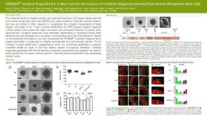 科学海报STEMdiff™ Cerebral Organoid Kit: A New Tool for the Culture of 3D Brain Organoids Derived from hPSCs
 沪公网安备31010102008431号