Wattanapanitch M et al. (SEP 2014)
PloS one 9 9 e106952
Dual small-molecule targeting of SMAD signaling stimulates human induced pluripotent stem cells toward neural lineages.
Incurable neurological disorders such as Parkinson's disease (PD),Huntington's disease (HD),and Alzheimer's disease (AD) are very common and can be life-threatening because of their progressive disease symptoms with limited treatment options. To provide an alternative renewable cell source for cell-based transplantation and as study models for neurological diseases,we generated induced pluripotent stem cells (iPSCs) from human dermal fibroblasts (HDFs) and then differentiated them into neural progenitor cells (NPCs) and mature neurons by dual SMAD signaling inhibitors. Reprogramming efficiency was improved by supplementing the histone deacethylase inhibitor,valproic acid (VPA),and inhibitor of p160-Rho associated coiled-coil kinase (ROCK),Y-27632,after retroviral transduction. We obtained a number of iPS colonies that shared similar characteristics with human embryonic stem cells in terms of their morphology,cell surface antigens,pluripotency-associated gene and protein expressions as well as their in vitro and in vivo differentiation potentials. After treatment with Noggin and SB431542,inhibitors of the SMAD signaling pathway,HDF-iPSCs demonstrated rapid and efficient differentiation into neural lineages. Six days after neural induction,neuroepithelial cells (NEPCs) were observed in the adherent monolayer culture,which had the ability to differentiate further into NPCs and neurons,as characterized by their morphology and the expression of neuron-specific transcripts and proteins. We propose that our study may be applied to generate neurological disease patient-specific iPSCs allowing better understanding of disease pathogenesis and drug sensitivity assays.
View Publication
产品号#:
05850
05857
05870
05875
07923
85850
85857
85870
85875
产品名:
Dispase (1 U/mL)
mTeSR™1
mTeSR™1
Busskamp V et al. (NOV 2014)
Molecular systems biology 10 11 760
Rapid neurogenesis through transcriptional activation in human stem cells.
Advances in cellular reprogramming and stem cell differentiation now enable ex vivo studies of human neuronal differentiation. However,it remains challenging to elucidate the underlying regulatory programs because differentiation protocols are laborious and often result in low neuron yields. Here,we overexpressed two Neurogenin transcription factors in human-induced pluripotent stem cells and obtained neurons with bipolar morphology in 4 days,at greater than 90% purity. The high purity enabled mRNA and microRNA expression profiling during neurogenesis,thus revealing the genetic programs involved in the rapid transition from stem cell to neuron. The resulting cells exhibited transcriptional,morphological and functional signatures of differentiated neurons,with greatest transcriptional similarity to prenatal human brain samples. Our analysis revealed a network of key transcription factors and microRNAs that promoted loss of pluripotency and rapid neurogenesis via progenitor states. Perturbations of key transcription factors affected homogeneity and phenotypic properties of the resulting neurons,suggesting that a systems-level view of the molecular biology of differentiation may guide subsequent manipulation of human stem cells to rapidly obtain diverse neuronal types.
View Publication
产品号#:
05854
05855
05850
05857
05870
05875
85850
85857
85870
85875
产品名:
mFreSR™
mFreSR™
mTeSR™1
mTeSR™1
Devlin A-C et al. (JAN 2015)
Nature Communications 6 1--12
Human iPSC-derived motoneurons harbouring TARDBP or C9ORF72 ALS mutations are dysfunctional despite maintaining viability
Crook JM et al. (MAR 2015)
Expert review of neurotherapeutics 15 3 295--304
The potential of induced pluripotent stem cells in models of neurological disorders: implications on future therapy.
There is an urgent need for new and advanced approaches to modeling the pathological mechanisms of complex human neurological disorders. This is underscored by the decline in pharmaceutical research and development efficiency resulting in a relative decrease in new drug launches in the last several decades. Induced pluripotent stem cells represent a new tool to overcome many of the shortcomings of conventional methods,enabling live human neural cell modeling of complex conditions relating to aberrant neurodevelopment,such as schizophrenia,epilepsy and autism as well as age-associated neurodegeneration. This review considers the current status of induced pluripotent stem cell-based modeling of neurological disorders,canvassing proven and putative advantages,current constraints,and future prospects of next-generation culture systems for biomedical research and translation.
View Publication
产品号#:
05850
05857
05870
05875
85850
85857
85870
85875
产品名:
mTeSR™1
mTeSR™1
Merkle FT et al. (FEB 2015)
Development (Cambridge,England) 142 4 633--643
Generation of neuropeptidergic hypothalamic neurons from human pluripotent stem cells.
Hypothalamic neurons orchestrate many essential physiological and behavioral processes via secreted neuropeptides,and are relevant to human diseases such as obesity,narcolepsy and infertility. We report the differentiation of human pluripotent stem cells into many of the major types of neuropeptidergic hypothalamic neurons,including those producing pro-opiolemelanocortin,agouti-related peptide,hypocretin/orexin,melanin-concentrating hormone,oxytocin,arginine vasopressin,corticotropin-releasing hormone (CRH) or thyrotropin-releasing hormone. Hypothalamic neurons can be generated using a 'self-patterning' strategy that yields a broad array of cell types,or via a more reproducible directed differentiation approach. Stem cell-derived human hypothalamic neurons share characteristic morphological properties and gene expression patterns with their counterparts in vivo,and are able to integrate into the mouse brain. These neurons could form the basis of cellular models,chemical screens or cellular therapies to study and treat common human diseases.
View Publication
产品号#:
05850
05857
05870
05875
85850
85857
85870
85875
产品名:
mTeSR™1
mTeSR™1
Su CTE et al. (FEB 2015)
Journal of visualized experiments : JoVE 96 1--9
An Optogenetic Approach for Assessing Formation of Neuronal Connections in a Co-culture System.
Here we describe a protocol to generate a co-culture consisting of 2 different neuronal populations. Induced pluripotent stem cells (iPSCs) are reprogrammed from human fibroblasts using episomal vectors. Colonies of iPSCs can be observed 30 days after initiation of fibroblast reprogramming. Pluripotent colonies are manually picked and grown in neural induction medium to permit differentiation into neural progenitor cells (NPCs). iPSCs rapidly convert into neuroepithelial cells within 1 week and retain the capability to self-renew when maintained at a high culture density. Primary mouse NPCs are differentiated into astrocytes by exposure to a serum-containing medium for 7 days and form a monolayer upon which embryonic day 18 (E18) rat cortical neurons (transfected with channelrhodopsin-2 (ChR2)) are added. Human NPCs tagged with the fluorescent protein,tandem dimer Tomato (tdTomato),are then seeded onto the astrocyte/cortical neuron culture the following day and allowed to differentiate for 28 to 35 days. We demonstrate that this system forms synaptic connections between iPSC-derived neurons and cortical neurons,evident from an increase in the frequency of synaptic currents upon photostimulation of the cortical neurons. This co-culture system provides a novel platform for evaluating the ability of iPSC-derived neurons to create synaptic connections with other neuronal populations.
View Publication
产品号#:
05850
05857
05870
05875
85850
85857
85870
85875
产品名:
mTeSR™1
mTeSR™1
Gallegos-Cá et al. (AUG 2015)
Stem cells and development 24 16 1901--1911
For diseases of the brain,the pig (Sus scrofa) is increasingly being used as a model organism that shares many anatomical and biological similarities with humans. We report that pig induced pluripotent stem cells (iPSC) can recapitulate events in early mammalian neural development. Pig iPSC line (POU5F1(high)/SSEA4(low)) had a higher potential to form neural rosettes (NR) containing neuroepithelial cells than either POU5F1(low)/SSEA4(low) or POU5F1(low)/SSEA4(high) lines. Thus,POU5F1 and SSEA4 pluripotency marker profiles in starting porcine iPSC populations can predict their propensity to form more robust NR populations in culture. The NR were isolated and expanded in vitro,retaining their NR morphology and neuroepithelial molecular properties. These cells expressed anterior central nervous system fate markers OTX2 and GBX2 through at least seven passages,and responded to retinoic acid,promoting a more posterior fate (HOXB4+,OTX2-,and GBX2-). These findings offer insight into pig iPSC development,which parallels the human iPSC in both anterior and posterior neural cell fates. These in vitro similarities in early neural differentiation processes support the use of pig iPSC and differentiated neural cells as a cell therapy in allogeneic porcine neural injury and degeneration models,providing relevant translational data for eventual human neural cell therapies.
View Publication
产品号#:
05850
05857
05870
05875
07923
85850
85857
85870
85875
产品名:
Dispase (1 U/mL)
mTeSR™1
mTeSR™1
Miranda C et al. (OCT 2015)
Biotechnology Journal 10 10 1612--1624
Spatial and temporal control of cell aggregation efficiently directs human pluripotent stem cells towards neural commitment
3D suspension culture is generally considered a promising method to achieve efficient expansion and controlled differentiation of human pluripotent stem cells (hPSCs). In this work,we focused on developing an integrated culture platform for expansion and neural commitment of hPSCs into neural precursors using 3D suspension conditions and chemically-defined culture media. We evaluated different inoculation methodologies for hPSC expansion as 3D aggregates and characterized the resulting cultures in terms of aggregate size distribution. It was demonstrated that upon single-cell inoculation,after four days of culture,3D aggregates were composed of homogenous populations of hPSC and were characterized by an average diameter of 139 ± 26 μm,which was determined to be the optimal size to initiate neural commitment. Temporal analysis revealed that upon neural specification it is possible to maximize the percentage of neural precursor cells expressing the neural markers Sox1 and Pax6 after nine days of culture. These results highlight our ability to define a robust method for production of hPSC-derived neural precursors that minimizes processing steps and that constitutes a promising alternative to the traditional planar adherent culture system due to a high potential for scaling-up.
View Publication
产品号#:
05850
05857
05870
05875
27305
85850
85857
85870
85875
产品名:
mTeSR™1
mTeSR™1
Handel AE et al. (MAR 2016)
Human Molecular Genetics 25 5 989--1000
Assessing similarity to primary tissue and cortical layer identity in induced pluripotent stem cell-derived cortical neurons through single-cell transcriptomics
Induced pluripotent stem cell (iPSC)-derived cortical neurons potentially present a powerful new model to understand corticogenesis and neurological disease. Previous work has established that differentiation protocols can produce cortical neurons,but little has been done to characterize these at cellular resolution. In particular,it is unclear to what extent in vitro two-dimensional,relatively disordered culture conditions recapitulate the development of in vivo cortical layer identity. Single-cell multiplex reverse transcriptase-quantitative polymerase chain reaction (RT-qPCR) was used to interrogate the expression of genes previously implicated in cortical layer or phenotypic identity in individual cells. Totally,93.6% of single cells derived from iPSCs expressed genes indicative of neuronal identity. High proportions of single neurons derived from iPSCs expressed glutamatergic receptors and synaptic genes. And,68.4% of iPSC-derived neurons expressing at least one layer marker could be assigned to a laminar identity using canonical cortical layer marker genes. We compared single-cell RNA-seq of our iPSC-derived neurons to available single-cell RNA-seq data from human fetal and adult brain and found that iPSC-derived cortical neurons closely resembled primary fetal brain cells. Unexpectedly,a subpopulation of iPSC-derived neurons co-expressed canonical fetal deep and upper cortical layer markers. However,this appeared to be concordant with data from primary cells. Our results therefore provide reassurance that iPSC-derived cortical neurons are highly similar to primary cortical neurons at the level of single cells but suggest that current layer markers,although effective,may not be able to disambiguate cortical layer identity in all cells.
View Publication
产品号#:
05850
05857
05870
05875
85850
85857
85870
85875
产品名:
mTeSR™1
mTeSR™1
Katori S et al. (JUL 2009)
The Journal of neuroscience : the official journal of the Society for Neuroscience 29 29 9137--47
Protocadherin-alpha family is required for serotonergic projections to appropriately innervate target brain areas.
Serotonergic axons from the raphe nuclei in the brainstem project to every region of the brain,where they make connections through their extensive terminal arborizations. This serotonergic innervation contributes to various normal behaviors and psychiatric disorders. The protocadherin-alpha (Pcdha) family of clustered protocadherins consists of 14 cadherin-related molecules generated from a single gene cluster. We found that the Pcdhas were strongly expressed in the serotonergic neurons. To elucidate their roles,we examined serotonergic fibers in a mouse mutant (Pcdha(Delta CR/Delta CR)) lacking the Pcdha cytoplasmic region-encoding exons,which are common to the gene cluster. In the first week after birth,the distribution pattern of serotonergic fibers in Pcdha(Delta CR/Delta CR) mice was similar to wild-type,but by 3 weeks of age,when the serotonergic axonal termini complete their arborizations,the distribution of the projections was abnormal. In some target regions,notably the globus pallidus and substantia nigra,the normally even distribution of serotonin axonal terminals was,in the mutants,dense at the periphery of each region,but sparse in the center. In the stratum lacunosum-molecular of the hippocampus,the mutants showed denser serotonergic innervation than in wild-type,and in the dentate gyrus of the hippocampus and the caudate-putamen,the innervation was sparser. Together,the abnormalities suggested that Pcdha proteins are important in the late-stage maturation of serotonergic projections. Further examination of alternatively spliced exons encoding the cytoplasmic tail showed that the A-type (but not the B-type) cytoplasmic tail was essential for the normal development of serotonergic projections.
View Publication
产品号#:
03800
03801
03802
03803
03804
03805
03806
产品名:
ClonaCell™-HY杂交瘤试剂盒
ClonaCell™-HY培养基A
ClonaCell™-HY 培养基 B
ClonaCell™-HY 培养基 C
ClonaCell™-HY 培养基 D
ClonaCell™-HY 培养基 E
ClonaCell™-HY PEG
Dobie FA and Craig AM (JUL 2011)
The Journal of neuroscience : the official journal of the Society for Neuroscience 31 29 10481--93
Inhibitory synapse dynamics: coordinated presynaptic and postsynaptic mobility and the major contribution of recycled vesicles to new synapse formation.
Dynamics of GABAergic synaptic components have been studied previously over milliseconds to minutes,revealing mobility of postsynaptic scaffolds and receptors. Here we image inhibitory synapses containing fluorescently tagged postsynaptic scaffold Gephyrin,together with presynaptic vesicular GABA transporter (VGAT) or postsynaptic GABA(A) receptor γ2 subunit (GABA(A)Rγ2),over seconds to days in cultured rat hippocampal neurons,revealing modes of inhibitory synapse formation and remodeling. Entire synapses were mobile,translocating rapidly within a confined region and exhibiting greater nonstochastic motion over multihour periods. Presynaptic and postsynaptic components moved in unison,maintaining close apposition while translocating distances of several micrometers. An observed flux in the density of synaptic puncta partially resulted from the apparent merging and splitting of preexisting clusters. De novo formation of inhibitory synapses was observed,marked by the appearance of stably apposed Gephyrin and VGAT clusters at sites previously lacking either component. Coclustering of GABA(A)Rγ2 supports the identification of such new clusters as synapses. Nascent synapse formation occurred by gradual accumulation of components over several hours,with VGAT clustering preceding that of Gephyrin and GABA(A)Rγ2. Comparing VGAT labeling by active uptake of a luminal domain antibody with post hoc immunocytochemistry indicated that recycling vesicles from preexisting boutons significantly contribute to vesicle pools at the majority of new inhibitory synapses. Although new synapses formed primarily on dendrite shafts,some also formed on dendritic protrusions,without apparent interconversion. Altogether,the long-term imaging of GABAergic presynaptic and postsynaptic components reveals complex dynamics and perpetual remodeling with implications for mechanisms of assembly and synaptic integration.
View Publication

 EasySep™小鼠TIL(CD45)正选试剂盒


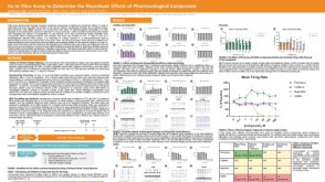 科学海报An In Vitro Assay to Determine the Neurotoxic Effects of Pharmacological Compounds
 沪公网安备31010102008431号