Sharma S et al. ( 2015)
Nature Communications 6 6881
APOBEC3A cytidine deaminase induces RNA editing in monocytes and macrophages
The extent,regulation and enzymatic basis of RNA editing by cytidine deamination are incompletely understood. Here we show that transcripts of hundreds of genes undergo site-specific CtextgreaterU RNA editing in macrophages during M1 polarization and in monocytes in response to hypoxia and interferons. This editing alters the amino acid sequences for scores of proteins,including many that are involved in pathogenesis of viral diseases. APOBEC3A,which is known to deaminate cytidines of single-stranded DNA and to inhibit viruses and retrotransposons,mediates this RNA editing. Amino acid residues of APOBEC3A that are known to be required for its DNA deamination and anti-retrotransposition activities were also found to affect its RNA deamination activity. Our study demonstrates the cellular RNA editing activity of a member of the APOBEC3 family of innate restriction factors and expands the understanding of CtextgreaterU RNA editing in mammals.
View Publication
产品号#:
19059
19059RF
产品名:
EasySep™人单核细胞富集试剂盒
RoboSep™ 人单核细胞富集试剂盒含滤芯吸头
Nudel I et al. (JAN 2011)
Journal of immunology (Baltimore,Md. : 1950) 186 2 891--900
Dendritic cells in distinct oral mucosal tissues engage different mechanisms to prime CD8+ T cells.
Although oral dendritic cells (DCs) were shown to induce cell-mediated immunity,the identity and function of the various oral DC subsets involved in this process is unclear. In this study,we examined the mechanisms used by DCs of the buccal mucosa and of the lining mucosa to elicit immunity. After plasmid DNA immunization,buccally immunized mice generated robust local and systemic CD8(+) T cell responses,whereas lower responses were seen by lining immunization. A delayed Ag presentation was monitored in vivo in both groups; yet,a more efficient presentation was mediated by buccal-derived DCs. Restricting transgene expression to CD11c(+) cells resulted in diminished CD8(+) T cell responses in both oral tissues,suggesting that immune induction is mediated mainly by cross-presentation. We then identified,in addition to the previously characterized Langerhans cells (LCs) and interstitial dendritic cells (iDCs),a third DC subset expressing the CD103(+) molecule,which represents an uncharacterized subset of oral iDCs expressing the langerin receptor (Ln(+)iDCs). Using Langerin-DTR mice,we demonstrated that whereas LCs and Ln(+)iDCs were dispensable for T cell induction in lining-immunized mice,LCs were essential for optimal CD8(+) T cell priming in the buccal mucosa. Buccal LCs,however,failed to directly present Ag to CD8(+) T cells,an activity that was mediated by buccal iDCs and Ln(+)iDCs. Taken together,our findings suggest that the mechanisms engaged by oral DCs to prime T cells vary between oral mucosal tissues,thus emphasizing the complexity of the oral immune network. Furthermore,we found a novel regulatory role for buccal LCs in potentiating CD8(+) T cell responses.
View Publication
产品号#:
19758
产品名:
Yang Q et al. (MAR 2011)
Blood 117 13 3529--38
E47 regulates hematopoietic stem cell proliferation and energetics but not myeloid lineage restriction.
The immune system is replenished by self-renewing hematopoietic stem cells (HSCs) that produce multipotent progenitors (MPPs) with little renewal capacity. E-proteins,the widely expressed basic helix-loop-helix transcription factors,contribute to HSC and MPP activity,but their specific functions remain undefined. Using quantitative in vivo and in vitro approaches,we show that E47 is dispensable for the short-term myeloid differentiation of HSCs but regulates their long-term capabilities. E47-deficient progenitors show competent myeloid production in short-term assays in vitro and in vivo. However,long-term myeloid and lymphoid differentiation is compromised because of a progressive loss of HSC self-renewal that is associated with diminished p21 expression and hyperproliferation. The activity of E47 is shown to be cell-intrinsic. Moreover,E47-deficient HSCs and MPPs have altered expression of genes associated with cellular energy metabolism,and the size of the MPP pool but not downstream lymphoid precursors in bone marrow or thymus is rescued in vivo by antioxidant. Together,these observations suggest a role for E47 in the tight control of HSC proliferation and energy metabolism,and demonstrate that E47 is not required for short-term myeloid differentiation.
View Publication
S. Balu et al. ( 2011)
The Journal of Immunology 186 3113-3119
A novel human IgA monoclonal antibody protects against tuberculosis
Abs have been shown to be protective in passive immunotherapy of tuberculous infection using mouse experimental models. In this study,we report on the properties of a novel human IgA1,constructed using a single-chain variable fragment clone (2E9),selected from an Ab phage library. The purified Ab monomer revealed high binding affinities for the mycobacterial ?-crystallin Ag and for the human Fc?RI (CD89) IgA receptor. Intranasal inoculations with 2E9IgA1 and recombinant mouse IFN-? significantly inhibited pulmonary H37Rv infection in mice transgenic for human CD89 but not in CD89-negative littermate controls,suggesting that binding to CD89 was necessary for the IgA-imparted passive protection. 2E9IgA1 added to human whole-blood or monocyte cultures inhibited luciferase-tagged H37Rv infection although not for all tested blood donors. Inhibition by 2E9IgA1 was synergistic with human rIFN-? in cultures of purified human monocytes but not in whole-blood cultures. The demonstration of the mandatory role of Fc?RI (CD89) for human IgA-mediated protection is important for understanding of the mechanisms involved and also for translation of this approach toward development of passive immunotherapy of tuberculosis.
View Publication
While a third of the world carries the burden of tuberculosis,disease control has been hindered by a lack of tools,including a rapid,point-of-care diagnostic and a protective vaccine. In many infectious diseases,antibodies (Abs) are powerful biomarkers and important immune mediators. However,in Mycobacterium tuberculosis (Mtb) infection,a discriminatory or protective role for humoral immunity remains unclear. Using an unbiased antibody profiling approach,we show that individuals with latent tuberculosis infection (Ltb) and active tuberculosis disease (Atb) have distinct Mtb-specific humoral responses,such that Ltb infection is associated with unique Ab Fc functional profiles,selective binding to FcγRIII,and distinct Ab glycosylation patterns. Moreover,compared to Abs from Atb,Abs from Ltb drove enhanced phagolysosomal maturation,inflammasome activation,and,most importantly,macrophage killing of intracellular Mtb. Combined,these data point to a potential role for Fc-mediated Ab effector functions,tuned via differential glycosylation,in Mtb control.
View Publication
产品号#:
18085
18085RF
18058
18058RF
15025
15065
产品名:
RosetteSep™人NK细胞富集抗体混合物
RosetteSep™人NK细胞富集抗体混合物
Figueroa G et al. (OCT 2016)
Journal of visualized experiments : JoVE 116
Characterization of Human Monocyte-derived Dendritic Cells by Imaging Flow Cytometry: A Comparison between Two Monocyte Isolation Protocols.
Dendritic cells (DCs) are antigen presenting cells of the immune system that play a crucial role in lymphocyte responses,host defense mechanisms,and pathogenesis of inflammation. Isolation and study of DCs have been important in biological research because of their distinctive features. Although they are essential key mediators of the immune system,DCs are very rare in blood,accounting for approximately 0.1 - 1% of total blood mononuclear cells. Therefore,alternatives for isolation methods rely on the differentiation of DCs from monocytes isolated from peripheral blood mononuclear cells (PBMCs). The utilization of proper isolation techniques that combine simplicity,affordability,high purity,and high yield of cells is imperative to consider. In the current study,two distinct methods for the generation of DCs will be compared. Monocytes were selected by adherence or negatively enriched using magnetic separation procedure followed by differentiation into DCs with IL-4 and GM-CSF. Monocyte and MDDC viability,proliferation,and phenotype were assessed using viability dyes,MTT assay,and CD11c/ CD14 surface marker analysis by imaging flow cytometry. Although the magnetic separation method yielded a significant higher percentage of monocytes with higher proliferative capacity when compared to the adhesion method,the findings have demonstrated the ability of both techniques to simultaneously generate monocytes that are capable of proliferating and differentiating into viable CD11c+ MDDCs after seven days in culture. Both methods yielded textgreater 70% CD11c+ MDDCs. Therefore,our results provide insights that contribute to the development of reliable methods for isolation and characterization of human DCs.
View Publication
产品号#:
19059
19059RF
产品名:
EasySep™人单核细胞富集试剂盒
RoboSep™ 人单核细胞富集试剂盒含滤芯吸头
Li R et al. (NOV 2016)
Cancer research
Macrophage-secreted TNFα and TGFβ1 Influence Migration Speed and Persistence of Cancer Cells in 3D Tissue Culture via Independent Pathways.
The ability of a cancer cell to migrate through the dense extracellular matrix (ECM) within and surrounding the solid tumor is a critical determinant of metastasis. Macrophages enhance invasion and metastasis in the tumor microenvironment but the basis for their effects are not fully understood. Using a microfluidic 3D cell migration assay,we found that the presence of macrophages enhanced the speed and persistence of cancer cell migration through a 3D extracellular matrix in a matrix metalloproteinases (MMP)-dependent fashion. Mechanistic investigations revealed that macrophage-released TNFα and TGFβ1 mediated the observed behaviors by two distinct pathways. These factors synergistically enhanced migration persistence through a synergistic induction of NF-κB-dependent MMP1 expression in cancer cells. In contrast,macrophage-released TGFβ1 enhanced migration speed primarily by inducing MT1-MMP expression. Taken together,our results reveal new insights into how macrophages enhance cancer cell metastasis,and they identify TNFα and TGFβ1 dual blockade as an anti-metastatic strategy in solid tumors.
View Publication

 EasySep™小鼠TIL(CD45)正选试剂盒


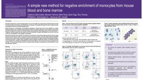 科学海报Method for Negative Enrichment of Monocytes from Mouse Blood and Bone Marrow
 挂图Antigen Processing and Presentation Overview of the mechanisms by which antigens are processed and presented to T cells
 沪公网安备31010102008431号