Harwood NMK et al. (MAR 2016)
Journal of leukocyte biology 99 3 495--503
HCV-infected cells and differentiation increase monocyte immunoregulatory galectin-9 production.
The lectin galectin-9 may help establish and maintain chronic hepatitis C virus infection. Galectin-9 is elevated in the liver and sera of hepatitis C virus patients,induces apoptosis of hepatitis C virus-specific T cells,and increases inhibitory regulatory T cells. Kupffer cells stain strongly for galectin-9 protein in hepatitis C virus patients. In the current study,we determined stimuli that induce galectin-9 production by monocytes and macrophages in hepatitis C virus infection. With the use of real-time PCR and flow cytometry,we analyzed galectin-9 mRNA and protein from human monocytes cocultured with hepatitis C virus-infected cells or noninfectious hepatitis C virus subgenomic replicon cells. We focused on finding the stimuli for galectin-9 production. Additionally,we measured galectin-9 during monocyte-to-macrophage maturation. Finally,we examined galectin-9 in peripheral monocytes from hepatitis C virus patients using flow cytometry. Galectin-9 mRNA increased 8-fold when primary monocytes were exposed to hepatitis C virus--infected cells. Maximum induction required proximity or contact and did not require IFN-γ or hepatitis C virus virions. Coculture of monocytes with subgenomic replicon cells increased galectin-9 5-fold,and purified exosomes from infected cells stimulated galectin-9 production. Stimulation of monocyte TLR3,-7,and -8 increased galectin-9 production. Differentiation of monocytes to macrophages increased galectin-9,and nonclassic monocytes from hepatitis C virus patients had the highest levels of galectin-9. Hepatitis C virus-infected cells stimulated monocytes to produce galectin-9 in close proximity,possibly,in part,as a result of exosomes and endosomal TLRs. Differentiation of monocytes to macrophages increased galectin-9. Nonclassic monocytes from hepatitis C virus patients express the highest galectin-9 levels,suggesting they may contribute to elevated galectin-9 and adaptive immune inhibition in hepatitis C virus infection.
View Publication
产品号#:
19059
19059RF
产品名:
EasySep™人单核细胞富集试剂盒
RoboSep™ 人单核细胞富集试剂盒含滤芯吸头
Borsa M et al. ( 2015)
The Virology Journal 12 77
HIV infection and antiretroviral therapy lead to unfolded protein response activation
BACKGROUND: The unfolded protein response (UPR) is one of the pathways triggered to ensure quality control of the proteins assembled in the endoplasmic reticulum (ER) when cell homeostasis is compromised. This mechanism is primarily composed of three transmembrane proteins serving as stress sensors: PKR-like ER kinase (PERK),activating transcription factor 6 (ATF6),and inositol-requiring enzyme 1 (IRE1). These three proteins' synergic action elicits translation and transcriptional downstream pathways,leading to less protein production and activating genes that encode important proteins in folding processes,including chaperones. Previous reports showed that viruses have evolved mechanisms to curtail or customize this UPR signaling for their own benefit. However,HIV infection's effect on the UPR has scarcely been investigated. METHODS: This work investigated UPR modulation by HIV infection by assessing UPR-related protein expression under in vitro and in vivo conditions via Western blotting. Antiretroviral (ARV) drugs' influence on this stress response was also considered. RESULTS: In in vitro and in vivo analyses,our results confirm that HIV infection activates stress-response components and that ARV therapy contributes to changes in the UPR's activation profile. CONCLUSIONS: This is the first report showing UPR-related protein expression in HIV target cells derived directly from HIV-infected patients receiving different ARV therapies. Thus,two mechanisms may occur simultaneously: interference by HIV itself and the ARV drugs' pharmacological effects as UPR activators. New evidence of how HIV modulates the UPR to enhance its own replication and secure infection success is also presented.
View Publication
产品号#:
15022
15062
15028
15068
产品名:
RosetteSep™人CD4+ T细胞富集抗体混合物
RosetteSep™人CD4+ T细胞富集抗体混合物
RosetteSep™人单核细胞富集抗体混合物
RosetteSep™人单核细胞富集抗体混合物
Currie KS et al. (MAY 2014)
Journal of medicinal chemistry 57 9 3856--73
Discovery of GS-9973, a selective and orally efficacious inhibitor of spleen tyrosine kinase.
Spleen tyrosine kinase (Syk) is an attractive drug target in autoimmune,inflammatory,and oncology disease indications. The most advanced Syk inhibitor,R406,1 (or its prodrug form fostamatinib,2),has shown efficacy in multiple therapeutic indications,but its clinical progress has been hampered by dose-limiting adverse effects that have been attributed,at least in part,to the off-target activities of 1. It is expected that a more selective Syk inhibitor would provide a greater therapeutic window. Herein we report the discovery and optimization of a novel series of imidazo[1,2-a]pyrazine Syk inhibitors. This work culminated in the identification of GS-9973,68,a highly selective and orally efficacious Syk inhibitor which is currently undergoing clinical evaluation for autoimmune and oncology indications.
View Publication
产品号#:
70034
70023
70023.1
200-0167
200-0166
产品名:
冻存的人外周血单核细胞
冻存的人外周血B细胞
冻存的人外周血B细胞
人外周血单核细胞,冷冻
人外周血单核细胞,冷冻
Christopher MJ et al. (FEB 2011)
The Journal of experimental medicine 208 2 251--60
Expression of the G-CSF receptor in monocytic cells is sufficient to mediate hematopoietic progenitor mobilization by G-CSF in mice.
Granulocyte colony-stimulating factor (G-CSF),the prototypical mobilizing cytokine,induces hematopoietic stem and progenitor cell (HSPC) mobilization from the bone marrow in a cell-nonautonomous fashion. This process is mediated,in part,through suppression of osteoblasts and disruption of CXCR4/CXCL12 signaling. The cellular targets of G-CSF that initiate the mobilization cascade have not been identified. We use mixed G-CSF receptor (G-CSFR)-deficient bone marrow chimeras to show that G-CSF-induced mobilization of HSPCs correlates poorly with the number of wild-type neutrophils. We generated transgenic mice in which expression of the G-CSFR is restricted to cells of the monocytic lineage. G-CSF-induced HSPC mobilization,osteoblast suppression,and inhibition of CXCL12 expression in the bone marrow of these transgenic mice are intact,demonstrating that G-CSFR signals in monocytic cells are sufficient to induce HSPC mobilization. Moreover,G-CSF treatment of wild-type mice is associated with marked loss of monocytic cells in the bone marrow. Finally,we show that bone marrow macrophages produce factors that support the growth and/or survival of osteoblasts in vitro. Together,these data suggest a model in which G-CSFR signals in bone marrow monocytic cells inhibit the production of trophic factors required for osteoblast lineage cell maintenance,ultimately leading to HSPC mobilization.
View Publication
Xu H et al. (JUL 2016)
Organic & biomolecular chemistry 14 26 6179--83
Cellular thermal shift and clickable chemical probe assays for the determination of drug-target engagement in live cells.
Proof of drug-target engagement in physiologically-relevant contexts is a key pillar of successful therapeutic target validation. We developed two orthogonal technologies,the cellular thermal shift assay (CETSA) and a covalent chemical probe reporter approach (harnessing sulfonyl fluoride tyrosine labeling and subsequent click chemistry) to measure the occupancy of the mRNA-decapping scavenger enzyme DcpS by a small molecule inhibitor in live cells. Enzyme affinity determined using isothermal dose response fingerprinting (ITDRFCETSA) and the concentration required to occupy 50% of the enzyme (OC50) using the chemical probe reporter assay were very similar. In this case,the chemical probe method worked well due to the long offset kinetics of the reversible inhibitor (determined using a fluorescent dye-tagged probe). This work suggests that CETSA could become the first choice assay to determine in-cell target engagement due to its simplicity.
View Publication
Application of the pMHC Array to Characterise Tumour Antigen Specific T Cell Populations in Leukaemia Patients at Disease Diagnosis.
Immunotherapy treatments for cancer are becoming increasingly successful,however to further improve our understanding of the T-cell recognition involved in effective responses and to encourage moves towards the development of personalised treatments for leukaemia immunotherapy,precise antigenic targets in individual patients have been identified. Cellular arrays using peptide-MHC (pMHC) tetramers allow the simultaneous detection of different antigen specific T-cell populations naturally circulating in patients and normal donors. We have developed the pMHC array to detect CD8+ T-cell populations in leukaemia patients that recognise epitopes within viral antigens (cytomegalovirus (CMV) and influenza (Flu)) and leukaemia antigens (including Per Arnt Sim domain 1 (PASD1),MelanA,Wilms' Tumour (WT1) and tyrosinase). We show that the pMHC array is at least as sensitive as flow cytometry and has the potential to rapidly identify more than 40 specific T-cell populations in a small sample of T-cells (0.8-1.4 x 10(6)). Fourteen of the twenty-six acute myeloid leukaemia (AML) patients analysed had T cells that recognised tumour antigen epitopes,and eight of these recognised PASD1 epitopes. Other tumour epitopes recognised were MelanA (n = 3),tyrosinase (n = 3) and WT1(126-134) (n = 1). One of the seven acute lymphocytic leukaemia (ALL) patients analysed had T cells that recognised the MUC1(950-958) epitope. In the future the pMHC array may be used provide point of care T-cell analyses,predict patient response to conventional therapy and direct personalised immunotherapy for patients.
View Publication

 EasySep™小鼠TIL(CD45)正选试剂盒


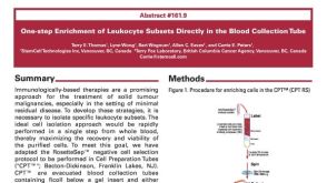 科学海报One-Step Enrichment of Leukocyte Subsets Directly in the Blood Collection Tube


 沪公网安备31010102008431号