Currie KS et al. (MAY 2014)
Journal of medicinal chemistry 57 9 3856--73
Discovery of GS-9973, a selective and orally efficacious inhibitor of spleen tyrosine kinase.
Spleen tyrosine kinase (Syk) is an attractive drug target in autoimmune,inflammatory,and oncology disease indications. The most advanced Syk inhibitor,R406,1 (or its prodrug form fostamatinib,2),has shown efficacy in multiple therapeutic indications,but its clinical progress has been hampered by dose-limiting adverse effects that have been attributed,at least in part,to the off-target activities of 1. It is expected that a more selective Syk inhibitor would provide a greater therapeutic window. Herein we report the discovery and optimization of a novel series of imidazo[1,2-a]pyrazine Syk inhibitors. This work culminated in the identification of GS-9973,68,a highly selective and orally efficacious Syk inhibitor which is currently undergoing clinical evaluation for autoimmune and oncology indications.
View Publication
Xu H et al. (JUL 2016)
Organic & biomolecular chemistry 14 26 6179--83
Cellular thermal shift and clickable chemical probe assays for the determination of drug-target engagement in live cells.
Proof of drug-target engagement in physiologically-relevant contexts is a key pillar of successful therapeutic target validation. We developed two orthogonal technologies,the cellular thermal shift assay (CETSA) and a covalent chemical probe reporter approach (harnessing sulfonyl fluoride tyrosine labeling and subsequent click chemistry) to measure the occupancy of the mRNA-decapping scavenger enzyme DcpS by a small molecule inhibitor in live cells. Enzyme affinity determined using isothermal dose response fingerprinting (ITDRFCETSA) and the concentration required to occupy 50% of the enzyme (OC50) using the chemical probe reporter assay were very similar. In this case,the chemical probe method worked well due to the long offset kinetics of the reversible inhibitor (determined using a fluorescent dye-tagged probe). This work suggests that CETSA could become the first choice assay to determine in-cell target engagement due to its simplicity.
View Publication
产品号#:
70025
70025.1
70025.2
70025.3
70047
70047.1
70047.2
70048
70048.1
70048.2
产品名:
冻存的人外周血单个核细胞
冻存的人外周血单个核细胞
冻存的人外周血单个核细胞
冻存的人外周血单个核细胞
Christopher MJ et al. (FEB 2011)
The Journal of experimental medicine 208 2 251--60
Expression of the G-CSF receptor in monocytic cells is sufficient to mediate hematopoietic progenitor mobilization by G-CSF in mice.
Granulocyte colony-stimulating factor (G-CSF),the prototypical mobilizing cytokine,induces hematopoietic stem and progenitor cell (HSPC) mobilization from the bone marrow in a cell-nonautonomous fashion. This process is mediated,in part,through suppression of osteoblasts and disruption of CXCR4/CXCL12 signaling. The cellular targets of G-CSF that initiate the mobilization cascade have not been identified. We use mixed G-CSF receptor (G-CSFR)-deficient bone marrow chimeras to show that G-CSF-induced mobilization of HSPCs correlates poorly with the number of wild-type neutrophils. We generated transgenic mice in which expression of the G-CSFR is restricted to cells of the monocytic lineage. G-CSF-induced HSPC mobilization,osteoblast suppression,and inhibition of CXCL12 expression in the bone marrow of these transgenic mice are intact,demonstrating that G-CSFR signals in monocytic cells are sufficient to induce HSPC mobilization. Moreover,G-CSF treatment of wild-type mice is associated with marked loss of monocytic cells in the bone marrow. Finally,we show that bone marrow macrophages produce factors that support the growth and/or survival of osteoblasts in vitro. Together,these data suggest a model in which G-CSFR signals in bone marrow monocytic cells inhibit the production of trophic factors required for osteoblast lineage cell maintenance,ultimately leading to HSPC mobilization.
View Publication
Neutrophil survival and c-kit(+)-progenitor proliferation in Staphylococcus aureus-infected skin wounds promote resolution.
Polymorphonuclear neutrophils (PMNs) are critical for the formation,maintenance,and resolution of bacterial abscesses. However,the mechanisms that regulate PMN survival and proliferation during the evolution of an abscess are not well defined. Using a mouse model of Staphylococcus aureus abscess formation within a cutaneous wound,combined with real-time imaging of genetically tagged PMNs,we observed that a high bacterial burden elicited a sustained mobilization of PMNs from the bone marrow to the infected wound,where their lifespan was markedly extended. A continuous rise in wound PMN number,which was not accounted for by trafficking from the bone marrow or by prolonged survival,was correlated with the homing of c-kit(+)-progenitor cells from the blood to the wound,where they proliferated and formed mature PMNs. Furthermore,by blocking their recruitment with an antibody to c-kit,which severely limited the proliferation of mature PMNs in the wound and shortened mouse survival,we confirmed that progenitor cells are not only important contributors to PMN expansion in the wound,but are also functionally important for immune protection. We conclude that the abscess environment provides a niche capable of regulating PMN survival and local proliferation of bone marrow-derived c-kit(+)-progenitor cells.
View Publication
产品号#:
03434
03444
产品名:
MethoCult™ GF M3434
MethoCult™ GF M3434
Harwood NMK et al. (MAR 2016)
Journal of leukocyte biology 99 3 495--503
HCV-infected cells and differentiation increase monocyte immunoregulatory galectin-9 production.
The lectin galectin-9 may help establish and maintain chronic hepatitis C virus infection. Galectin-9 is elevated in the liver and sera of hepatitis C virus patients,induces apoptosis of hepatitis C virus-specific T cells,and increases inhibitory regulatory T cells. Kupffer cells stain strongly for galectin-9 protein in hepatitis C virus patients. In the current study,we determined stimuli that induce galectin-9 production by monocytes and macrophages in hepatitis C virus infection. With the use of real-time PCR and flow cytometry,we analyzed galectin-9 mRNA and protein from human monocytes cocultured with hepatitis C virus-infected cells or noninfectious hepatitis C virus subgenomic replicon cells. We focused on finding the stimuli for galectin-9 production. Additionally,we measured galectin-9 during monocyte-to-macrophage maturation. Finally,we examined galectin-9 in peripheral monocytes from hepatitis C virus patients using flow cytometry. Galectin-9 mRNA increased 8-fold when primary monocytes were exposed to hepatitis C virus--infected cells. Maximum induction required proximity or contact and did not require IFN-γ or hepatitis C virus virions. Coculture of monocytes with subgenomic replicon cells increased galectin-9 5-fold,and purified exosomes from infected cells stimulated galectin-9 production. Stimulation of monocyte TLR3,-7,and -8 increased galectin-9 production. Differentiation of monocytes to macrophages increased galectin-9,and nonclassic monocytes from hepatitis C virus patients had the highest levels of galectin-9. Hepatitis C virus-infected cells stimulated monocytes to produce galectin-9 in close proximity,possibly,in part,as a result of exosomes and endosomal TLRs. Differentiation of monocytes to macrophages increased galectin-9. Nonclassic monocytes from hepatitis C virus patients express the highest galectin-9 levels,suggesting they may contribute to elevated galectin-9 and adaptive immune inhibition in hepatitis C virus infection.
View Publication

 EasySep™小鼠TIL(CD45)正选试剂盒


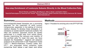 科学海报One-Step Enrichment of Leukocyte Subsets Directly in the Blood Collection Tube


 沪公网安备31010102008431号