Harwood NMK et al. (MAR 2016)
Journal of leukocyte biology 99 3 495--503
HCV-infected cells and differentiation increase monocyte immunoregulatory galectin-9 production.
The lectin galectin-9 may help establish and maintain chronic hepatitis C virus infection. Galectin-9 is elevated in the liver and sera of hepatitis C virus patients,induces apoptosis of hepatitis C virus-specific T cells,and increases inhibitory regulatory T cells. Kupffer cells stain strongly for galectin-9 protein in hepatitis C virus patients. In the current study,we determined stimuli that induce galectin-9 production by monocytes and macrophages in hepatitis C virus infection. With the use of real-time PCR and flow cytometry,we analyzed galectin-9 mRNA and protein from human monocytes cocultured with hepatitis C virus-infected cells or noninfectious hepatitis C virus subgenomic replicon cells. We focused on finding the stimuli for galectin-9 production. Additionally,we measured galectin-9 during monocyte-to-macrophage maturation. Finally,we examined galectin-9 in peripheral monocytes from hepatitis C virus patients using flow cytometry. Galectin-9 mRNA increased 8-fold when primary monocytes were exposed to hepatitis C virus--infected cells. Maximum induction required proximity or contact and did not require IFN-γ or hepatitis C virus virions. Coculture of monocytes with subgenomic replicon cells increased galectin-9 5-fold,and purified exosomes from infected cells stimulated galectin-9 production. Stimulation of monocyte TLR3,-7,and -8 increased galectin-9 production. Differentiation of monocytes to macrophages increased galectin-9,and nonclassic monocytes from hepatitis C virus patients had the highest levels of galectin-9. Hepatitis C virus-infected cells stimulated monocytes to produce galectin-9 in close proximity,possibly,in part,as a result of exosomes and endosomal TLRs. Differentiation of monocytes to macrophages increased galectin-9. Nonclassic monocytes from hepatitis C virus patients express the highest galectin-9 levels,suggesting they may contribute to elevated galectin-9 and adaptive immune inhibition in hepatitis C virus infection.
View Publication
产品号#:
19059
19059RF
产品名:
EasySep™人单核细胞富集试剂盒
RoboSep™ 人单核细胞富集试剂盒含滤芯吸头
Borsa M et al. ( 2015)
The Virology Journal 12 77
HIV infection and antiretroviral therapy lead to unfolded protein response activation
BACKGROUND: The unfolded protein response (UPR) is one of the pathways triggered to ensure quality control of the proteins assembled in the endoplasmic reticulum (ER) when cell homeostasis is compromised. This mechanism is primarily composed of three transmembrane proteins serving as stress sensors: PKR-like ER kinase (PERK),activating transcription factor 6 (ATF6),and inositol-requiring enzyme 1 (IRE1). These three proteins' synergic action elicits translation and transcriptional downstream pathways,leading to less protein production and activating genes that encode important proteins in folding processes,including chaperones. Previous reports showed that viruses have evolved mechanisms to curtail or customize this UPR signaling for their own benefit. However,HIV infection's effect on the UPR has scarcely been investigated. METHODS: This work investigated UPR modulation by HIV infection by assessing UPR-related protein expression under in vitro and in vivo conditions via Western blotting. Antiretroviral (ARV) drugs' influence on this stress response was also considered. RESULTS: In in vitro and in vivo analyses,our results confirm that HIV infection activates stress-response components and that ARV therapy contributes to changes in the UPR's activation profile. CONCLUSIONS: This is the first report showing UPR-related protein expression in HIV target cells derived directly from HIV-infected patients receiving different ARV therapies. Thus,two mechanisms may occur simultaneously: interference by HIV itself and the ARV drugs' pharmacological effects as UPR activators. New evidence of how HIV modulates the UPR to enhance its own replication and secure infection success is also presented.
View Publication
产品号#:
15022
15062
15028
15068
产品名:
RosetteSep™人CD4+ T细胞富集抗体混合物
RosetteSep™人CD4+ T细胞富集抗体混合物
RosetteSep™人单核细胞富集抗体混合物
RosetteSep™人单核细胞富集抗体混合物
Currie KS et al. (MAY 2014)
Journal of medicinal chemistry 57 9 3856--73
Discovery of GS-9973, a selective and orally efficacious inhibitor of spleen tyrosine kinase.
Spleen tyrosine kinase (Syk) is an attractive drug target in autoimmune,inflammatory,and oncology disease indications. The most advanced Syk inhibitor,R406,1 (or its prodrug form fostamatinib,2),has shown efficacy in multiple therapeutic indications,but its clinical progress has been hampered by dose-limiting adverse effects that have been attributed,at least in part,to the off-target activities of 1. It is expected that a more selective Syk inhibitor would provide a greater therapeutic window. Herein we report the discovery and optimization of a novel series of imidazo[1,2-a]pyrazine Syk inhibitors. This work culminated in the identification of GS-9973,68,a highly selective and orally efficacious Syk inhibitor which is currently undergoing clinical evaluation for autoimmune and oncology indications.
View Publication
产品号#:
70034
70023
70023.1
200-0167
200-0166
产品名:
冻存的人外周血单核细胞
冻存的人外周血B细胞
冻存的人外周血B细胞
人外周血单核细胞,冷冻
人外周血单核细胞,冷冻
Carrera Silva EA et al. ( 2017)
Blood 130 17 1898--1902
CD207+CD1a+ cells circulate in pediatric patients with active Langerhans cell histiocytosis.
Langerhans cell histiocytosis (LCH) is a rare disease with an unknown etiology characterized by heterogeneous lesions containing CD207+CD1a+ cells that can arise in almost any tissue and cause significant morbidity and mortality. Precursors of pathological Langerhans cells have yet to be defined. Our aim was to identify circulating CD207+CD1a+ cells and their inducers in LCH. Expression of CD207 and CD1a in the blood myeloid compartment as well as thymic stromal lymphopoietin (TSLP) and transforming growth factor β (TGF-β) plasma levels were measured in 22 pediatric patients with active disease (AD) or nonactive disease (NAD). In patients with AD vs those with NAD,the myeloid compartment showed an increased CD11b (CD11bhigh plus CD11b+) fraction (39.7 ± 3.6 vs 18.6 ± 1.9),a higher percentage of circulating CD11bhighCD11c+CD207+ cells (44.5 ± 11.3 vs 3.2 ± 0.5),and the presence of CD11chighCD207+CD1a+ cells (25.0 ± 9.1 vs 2.3 ± 0.5). Blood CD207+CD1a+ cells were not observed in adult controls or umbilical cord. Increased TSLP and TGF-β levels were detected in patients with AD. Interestingly,plasma from patients with AD induces CD207 expression on CD14+ monocytes. We conclude that CD207+CD1a+ cells are circulating in patients with active LCH,and TSLP and TGF-β are potential drivers of Langerhans-like cells in vivo.
View Publication
Lambrianides A et al. (JUN 2010)
Journal of immunology (Baltimore,Md. : 1950) 184 12 6622--8
Effects of polyclonal IgG derived from patients with different clinical types of the antiphospholipid syndrome on monocyte signaling pathways.
A major mechanism of hypercoagulability in the antiphospholipid syndrome (APS) is antiphospholipid Ab-mediated upregulation of tissue factor (TF) on monocytes via activation of TLRs,p38 MAPK,and NF-kappaB pathways. We examined whether monocyte signaling pathways are differentially activated by IgG from patients with vascular thrombosis (VT) alone compared with IgG from patients with pregnancy morbidity (PM) alone. We purified IgG from 49 subjects. A human monocyte cell line and ex vivo healthy monocytes were treated with 100 microg/ml IgG for 6 h,and cell extracts were examined by immunoblot using Abs to p38 MAPK and NF-kappaB. To further investigate intracellular signaling pathways induced by these IgGs,specific inhibitors of p38 MAPK,NF-kappaB,TLR4,and TLR2 were used to determine their effect on TF activity. Only IgG from patients with VT but no PM (VT+/PM-) caused phosphorylation of NF-kappaBand p38 MAPK and upregulation of TF activity in monocytes. These effects were not seen with IgG from patients with PM alone (VT-/PM+),anti-phospholipid Ab-positive patients without APS,or healthy controls. TF upregulation caused by the VT+/PM- samples was reduced by inhibitors of p38 MAPK,NF-kappaB,and TLR4. The effects of VT+/PM- IgG on signaling and TF upregulation were concentrated in the fraction that bound beta-2-glycoprotein I. Our findings demonstrate that IgGs from patients with diverse clinical manifestations of APS have differential effects upon phosphorylation of NF-kappaB and p38 MAPK and TF activity that may be mediated by differential activation of TLR4.
View Publication
Application of the pMHC Array to Characterise Tumour Antigen Specific T Cell Populations in Leukaemia Patients at Disease Diagnosis.
Immunotherapy treatments for cancer are becoming increasingly successful,however to further improve our understanding of the T-cell recognition involved in effective responses and to encourage moves towards the development of personalised treatments for leukaemia immunotherapy,precise antigenic targets in individual patients have been identified. Cellular arrays using peptide-MHC (pMHC) tetramers allow the simultaneous detection of different antigen specific T-cell populations naturally circulating in patients and normal donors. We have developed the pMHC array to detect CD8+ T-cell populations in leukaemia patients that recognise epitopes within viral antigens (cytomegalovirus (CMV) and influenza (Flu)) and leukaemia antigens (including Per Arnt Sim domain 1 (PASD1),MelanA,Wilms' Tumour (WT1) and tyrosinase). We show that the pMHC array is at least as sensitive as flow cytometry and has the potential to rapidly identify more than 40 specific T-cell populations in a small sample of T-cells (0.8-1.4 x 10(6)). Fourteen of the twenty-six acute myeloid leukaemia (AML) patients analysed had T cells that recognised tumour antigen epitopes,and eight of these recognised PASD1 epitopes. Other tumour epitopes recognised were MelanA (n = 3),tyrosinase (n = 3) and WT1(126-134) (n = 1). One of the seven acute lymphocytic leukaemia (ALL) patients analysed had T cells that recognised the MUC1(950-958) epitope. In the future the pMHC array may be used provide point of care T-cell analyses,predict patient response to conventional therapy and direct personalised immunotherapy for patients.
View Publication

 EasySep™小鼠TIL(CD45)正选试剂盒


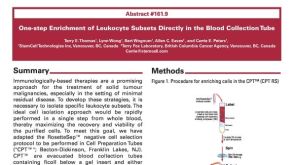 科学海报One-Step Enrichment of Leukocyte Subsets Directly in the Blood Collection Tube


 沪公网安备31010102008431号