Tomé et al. (AUG 2016)
The Journal of nutritional biochemistry 34 146--55
Hydroxytyrosol supplementation modulates the expression of miRNAs in rodents and in humans.
Dietary microRNAs (miRNAs) modulation could be important for health and wellbeing. Part of the healthful activities of polyphenols might be due to a modulation of miRNAs' expression. Among the most biologically active polyphenols,hydroxytyrosol (HT) has never been studied for its actions on miRNAs. We investigated whether HT could modulate the expression of miRNAs in vivo. We performed an unbiased intestinal miRNA screening in mice supplemented (for 8 weeks) with nutritionally relevant amounts of HT. HT modulated the expression of several miRNAs. Analysis of other tissues revealed consistent HT-induced modulation of only few miRNAs. Also,HT administration increased triglycerides levels. Acute treatment with HT and in vitro experiments provided mechanistic insights. The HT-induced expression of one miRNA was confirmed in healthy volunteers supplemented with HT in a randomized,double-blind and placebo-controlled trial. HT consumption affects specific miRNAs' expression in rodents and humans. Our findings suggest that the modulation of miRNAs' action through HT consumption might partially explain its healthful activities and might be pharmanutritionally exploited in current therapies targeting endogenous miRNAs. However,the effects of HT on triglycerides warrant further investigations.
View Publication
Increased Abundance of M Cells in the Gut Epithelium Dramatically Enhances Oral Prion Disease Susceptibility.
Many natural prion diseases of humans and animals are considered to be acquired through oral consumption of contaminated food or pasture. Determining the route by which prions establish host infection will identify the important factors that influence oral prion disease susceptibility and to which intervention strategies can be developed. After exposure,the early accumulation and replication of prions within small intestinal Peyer's patches is essential for the efficient spread of disease to the brain. To replicate within Peyer's patches,the prions must first cross the gut epithelium. M cells are specialised epithelial cells within the epithelia covering Peyer's patches that transcytose particulate antigens and microorganisms. M cell-development is dependent upon RANKL-RANK-signalling,and mice in which RANK is deleted only in the gut epithelium completely lack M cells. In the specific absence of M cells in these mice,the accumulation of prions within Peyer's patches and the spread of disease to the brain was blocked,demonstrating a critical role for M cells in the initial transfer of prions across the gut epithelium in order to establish host infection. Since pathogens,inflammatory stimuli and aging can modify M cell-density in the gut,these factors may also influence oral prion disease susceptibility. Mice were therefore treated with RANKL to enhance M cell density in the gut. We show that prion uptake from the gut lumen was enhanced in RANKL-treated mice,resulting in shortened survival times and increased disease susceptibility,equivalent to a 10-fold higher infectious titre of prions. Together these data demonstrate that M cells are the critical gatekeepers of oral prion infection,whose density in the gut epithelium directly limits or enhances disease susceptibility. Our data suggest that factors which alter M cell-density in the gut epithelium may be important risk factors which influence host susceptibility to orally acquired prion diseases.
View Publication
Alison MR et al. (DEC 2010)
The Journal of pathology 222 4 335--44
Finding cancer stem cells: are aldehyde dehydrogenases fit for purpose?
Despite many years of intensive effort,there is surprisingly little consensus on the most suitable markers with which to locate and isolate stem cells from adult tissues. By comparison,the study of cancer stem cells is still in its infancy; so,unsurprisingly,there is great uncertainty as to the identity of these cells. Stem cell markers can be broadly categorized into molecular determinants of self-renewal,clonogenicity,multipotentiality,adherence to the niche,and longevity. This review assesses the utility of recognizing cancer stem cells by virtue of high expression of aldehyde dehydrogenases (ALDHs),probably significant determinants of cell survival through their ability to detoxify many potentially cytotoxic molecules,and contributing to drug resistance. Antibodies are available against the ALDH enzyme family,but the vast majority of studies have used cell sorting techniques to enrich for cells expressing these enzymes. Live cells expressing high ALDH activity are usually identified by the ALDEFLUOR kit and sorted by fluorescence activated cell sorting (FACS). For many human tumours,but notably breast cancer,cell selection based upon ALDH activity appears to be a useful marker for enriching for cells with tumour-initiating activity (presumed cancer stem cells) in immunodeficient mice,and indeed the frequency of so-called ALDH(bri) cells in many tumours can be an independent prognostic indicator.
View Publication
产品号#:
01700
01705
01702
产品名:
ALDEFLUOR™ 试剂盒
ALDEFLUOR™ DEAB试剂, 1.5 mM, 1 mL
ALDEFLUOR™检测缓冲液
Taubert I et al. (APR 2011)
Cytotherapy 13 4 459--66
Characterization of hematopoietic stem cell subsets from patients with multiple myeloma after mobilization with plerixafor.
BACKGROUND AIMS: Previous studies have demonstrated that the combination of granulocyte-colony-stimulating factor (G-CSF) + plerixafor is more efficient in mobilizing CD34(+) hematopoietic stem cells (HSC) into the peripheral blood than G-CSF alone. In this study we analyzed the impact of adding plerixafor to G-CSF upon the mobilization of different HSC subsets. METHODS: We characterized the immunophenotype of HSC subsets isolated from the peripheral blood of eight patients with multiple myeloma (MM) before and after treatment with plerixafor. All patients were supposed to collect stem cells prior to high-dose chemotherapy and consecutive autologous stem cell transplantation,and therefore received front-line mobilization with 4 days of G-CSF followed by a single dose of plerixafor. Samples of peripheral blood were analyzed comparatively by flow cytometry directly before and 12 h after administration of plerixafor. RESULTS: The number of aldehyde dehydrogenase (ALDH)(bright) and CD34(+) cells was significantly higher after plerixafor treatment (1.2-5.0 and 1.5-6.0 times; both P textless 0.01) and an enrichment of the very primitive CD34(+) CD38(-) and ALDH(bright) CD34(+) CD38(-) HSC subsets was detectable. Additionally,two distinct ALDH(+) subsets could be clearly distinguished. The small ALDH(high) subset showed a higher number of CD34(+) CD38(-) cells in contrast to the total ALDH(bright) subpopulation and probably represented a very primitive subpopulation of HSC. CONCLUSIONS: A combined staining of ALDH,CD34 and CD38 might represent a powerful tool for the identification of a very rare and primitive hematopoietic stem cell subset. The addition of plerixafor mobilized not only more CD34(+) cells but was also able to increase the proportion of more primitive stem cell subsets.
View Publication
产品号#:
01700
01705
01702
产品名:
ALDEFLUOR™ 试剂盒
ALDEFLUOR™ DEAB试剂, 1.5 mM, 1 mL
ALDEFLUOR™检测缓冲液
Sullivan JP et al. (DEC 2010)
Cancer research 70 23 9937--48
Aldehyde dehydrogenase activity selects for lung adenocarcinoma stem cells dependent on notch signaling.
Aldehyde dehydrogenase (ALDH) is a candidate marker for lung cancer cells with stem cell-like properties. Immunohistochemical staining of a large panel of primary non-small cell lung cancer (NSCLC) samples for ALDH1A1,ALDH3A1,and CD133 revealed a significant correlation between ALDH1A1 (but not ALDH3A1 or CD133) expression and poor prognosis in patients including those with stage I and N0 disease. Flow cytometric analysis of a panel of lung cancer cell lines and patient tumors revealed that most NSCLCs contain a subpopulation of cells with elevated ALDH activity,and that this activity is associated with ALDH1A1 expression. Isolated ALDH(+) lung cancer cells were observed to be highly tumorigenic and clonogenic as well as capable of self-renewal compared with their ALDH(-) counterparts. Expression analysis of sorted cells revealed elevated Notch pathway transcript expression in ALDH(+) cells. Suppression of the Notch pathway by treatment with either a γ-secretase inhibitor or stable expression of shRNA against NOTCH3 resulted in a significant decrease in ALDH(+) lung cancer cells,commensurate with a reduction in tumor cell proliferation and clonogenicity. Taken together,these findings indicate that ALDH selects for a subpopulation of self-renewing NSCLC stem-like cells with increased tumorigenic potential,that NSCLCs harboring tumor cells with ALDH1A1 expression have inferior prognosis,and that ALDH1A1 and CD133 identify different tumor subpopulations. Therapeutic targeting of the Notch pathway reduces this ALDH(+) component,implicating Notch signaling in lung cancer stem cell maintenance.
View Publication

 EasySep™小鼠TIL(CD45)正选试剂盒


 实验方案Cryopreserving Hepatic Organoids Expanded in HepatiCult™ Organoid Growth Medium (Human)
 实验方案Recovery and Expansion of Hepatic Organoids Using HepatiCult™ Organoid Growth Medium
 实验方案Expansion of Hepatic Organoids via Single Cells Using HepatiCult™ Organoid Growth Medium
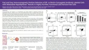 科学海报Positive Selection of PE- or Biotin-Conjugated Antibody Labeled Cells with Releasable Rapidspheres™

 沪公网安备31010102008431号