Chen Y et al. (JUL 2009)
Journal of immunology (Baltimore,Md. : 1950) 183 2 1346--59
Regulation of dendritic cells and macrophages by an anti-apoptotic cell natural antibody that suppresses TLR responses and inhibits inflammatory arthritis.
Although natural Abs (NAbs) are present from birth,little is known about what drives their selection and whether they have housekeeping functions. The prototypic T15-NAb,first identified because of its protective role in infection,is representative of a special type of NAb response that specifically recognizes and forms complexes with apoptotic cells and which promotes cell-corpse engulfment by phagocytes. We now show that this T15-NAb IgM-mediated clearance process is dependent on the recruitment of C1q and mannose-binding lectin,which have known immune modulatory activities that also provide eat me" signals for enhancing phagocytosis. Further investigation revealed that the addition of T15-NAb significantly suppressed in vitro LPS-induced TNF-alpha and IL-6 secretion by the macrophage-like cell line�
View Publication
产品号#:
09600
09650
产品名:
StemSpan™ SFEM
StemSpan™ SFEM
Costantini C et al. (JAN 2009)
Immunobiology 214 9-10 828--34
On the co-purification of 6-sulfo LacNAc(+) dendritic cells (slanDC) with NK cells enriched from human blood.
The ability of NK cells to directly recognize pathogens and be activated via Toll-like receptors (TLR) is increasingly recognized. Nevertheless,controversial results on the NK cell ability to be directly activated by lipopolysaccharide (LPS),the ligand of TLR4,have been recently reported. To start elucidating the reasons explaining the contrasting observations of the literature,we focused on the potential role of currently used NK cell purification procedures to condition putative NK cell responsiveness to LPS. To do so,human NK cells were isolated by negative selection,using three different commercial kits,to be comparatively evaluated for the production of IFNgamma in response to ultra-pure LPS and/or IL-2. Despite the lack of surface TLR4,we found that two out of the three NK cell-enriched populations released IFNgamma (and one of the two,IL-12p70 as well) in response to the LPS plus IL-2 combination,whereas the last one did not. However,the two LPS plus IL-2-responsive NK cell populations were found variably contaminated with 6-sulfo LacNAc(+) dendritic cells (slanDC),demonstrated responsible for triggering,via the production of IL-12p70 in response to LPS,the release of IFNgamma by IL-2-stimulated NK cells. Accordingly,slanDC depletion completely abrogated the capacity to produce both IL-12p70 and IFNgamma in response to LPS plus IL-2 by slanDC-containing NK cells. Taken together,our data uncover that two commercially available kits,specifically designed to isolate NK cells by negative selection,also co-purify variable amounts of slanDC. The latter cells may dramatically affect the outcome of experiments carried on to evaluate NK cell responsiveness to TLR agonists such as LPS.
View Publication
产品号#:
19055
19055RF
产品名:
EasySep™人NK细胞富集试剂盒
RoboSep™ 人NK细胞富集试剂盒含滤芯吸头
Guilliams M et al. (MAR 2010)
Blood 115 10 1958--68
Skin-draining lymph nodes contain dermis-derived CD103(-) dendritic cells that constitutively produce retinoic acid and induce Foxp3(+) regulatory T cells.
Small intestinal CD103(+) dendritic cells (DCs) have the selective ability to promote de novo generation of regulatory T cells via the production of retinoic acid (RA). Considering that aldehyde dehydrogenase (ALDH) activity controls the production of RA,we used a flow cytometry-based assay to measure ALDH activity at the single-cell level and to perform a comprehensive analysis of the RA-producing DC populations present in lymphoid and nonlymphoid mouse tissues. RA-producing DCs were primarily of the tissue-derived,migratory DC subtype and can be readily found in the skin and in the lungs as well as in their corresponding draining lymph nodes. The RA-producing skin-derived DCs were capable of triggering the generation of regulatory T cells,a finding demonstrating that the presence of RA-producing,tolerogenic DCs is not restricted to the intestinal tract as previously thought. Unexpectedly,the production of RA by skin DCs was restricted to CD103(-) DCs,indicating that CD103 expression does not constitute a universal" marker for RA-producing mouse DCs. Finally�
View Publication
产品号#:
01700
01705
01702
产品名:
ALDEFLUOR™ 试剂盒
ALDEFLUOR™ DEAB试剂, 1.5 mM, 1 mL
ALDEFLUOR™检测缓冲液
Krummen M et al. (JUL 2010)
Journal of leukocyte biology 88 1 189--99
Release of IL-12 by dendritic cells activated by TLR ligation is dependent on MyD88 signaling, whereas TRIF signaling is indispensable for TLR synergy.
Recently,it has been shown that certain combinations of TLR ligands act in synergy to induce the release of IL-12 by DCs. In this study,we sought to define the critical parameters underlying TLR synergy. Our data show that TLR ligands act synergistically if MyD88- and TRIF-dependent ligands are combined. TLR4 uses both of these adaptor molecules,thus activation via TLR4 proved to be a synergistic event on its own. TLR synergy did not affect all aspects of DC activation but enhanced primarily the release of certain cytokines,particularly IL-12,whereas the expression of costimulatory molecules remained unchanged. Consequently,synergistic activation of DC did not affect their ability to induce T cell proliferation but resulted in T(H)1-biased responses in vitro and in vivo. Furthermore,we examined the impact of TLR ligand combinations on primary DC in vitro but observed only modest effects with a combination of CpG + Poly (I:C). However,noticeable synergy in terms of IL-12 production by DCs was detectable in vivo after systemic administration of CpG + Poly (I:C). Finally,we show that synergy is partially dependent on IFNAR signaling but does not require the release of IFNs to the enviroment,suggesting an autocrine action of type I IFNs.
View Publication
产品号#:
18752
18752RF
21000
20119
20155
18758
18758RF
18768
18768RF
产品名:
RoboSep™- S
RoboSep™ 吸头组件抛光剂
RoboSep™分选管套装(9个塑料管)
Poulin LF et al. (JUN 2010)
The Journal of experimental medicine 207 6 1261--71
Characterization of human DNGR-1+ BDCA3+ leukocytes as putative equivalents of mouse CD8alpha+ dendritic cells.
In mouse,a subset of dendritic cells (DCs) known as CD8alpha+ DCs has emerged as an important player in the regulation of T cell responses and a promising target in vaccination strategies. However,translation into clinical protocols has been hampered by the failure to identify CD8alpha+ DCs in humans. Here,we characterize a population of human DCs that expresses DNGR-1 (CLEC9A) and high levels of BDCA3 and resembles mouse CD8alpha+ DCs in phenotype and function. We describe the presence of such cells in the spleens of humans and humanized mice and report on a protocol to generate them in vitro. Like mouse CD8alpha+ DCs,human DNGR-1+ BDCA3hi DCs express Necl2,CD207,BATF3,IRF8,and TLR3,but not CD11b,IRF4,TLR7,or (unlike CD8alpha+ DCs) TLR9. DNGR-1+ BDCA3hi DCs respond to poly I:C and agonists of TLR8,but not of TLR7,and produce interleukin (IL)-12 when given innate and T cell-derived signals. Notably,DNGR-1+ BDCA3+ DCs from in vitro cultures efficiently internalize material from dead cells and can cross-present exogenous antigens to CD8+ T cells upon treatment with poly I:C. The characterization of human DNGR-1+ BDCA3hi DCs and the ability to grow them in vitro opens the door for exploiting this subset in immunotherapy.
View Publication
产品号#:
09600
09650
产品名:
StemSpan™ SFEM
StemSpan™ SFEM
Poulsen C et al. (AUG 2015)
Toxicology letters 237 1 21--9
Differential cytotoxicity of long-chain bases for human oral gingival epithelial keratinocytes, oral fibroblasts, and dendritic cells.
Long-chain bases are present in the oral cavity. Previously we determined that sphingosine,dihydrosphingosine,and phytosphingosine have potent antimicrobial activity against oral pathogens. Here,we determined the cytotoxicities of long-chain bases for oral cells,an important step in considering their potential as antimicrobial agents for oral infections. This information would clearly help in establishing prophylactic or therapeutic doses. To assess this,human oral gingival epithelial (GE) keratinocytes,oral gingival fibroblasts (GF),and dendritic cells (DC) were exposed to 10.0-640.0 μM long-chain bases and glycerol monolaurate (GML). The effects of long-chain bases on cell metabolism (conversion of resazurin to resorufin),membrane permeability (uptake of propidium iodide or SYTOX-Green),release of cellular contents (LDH),and cell morphology (confocal microscopy) were all determined. GE keratinocytes were more resistant to long-chain bases as compared to GF and DC,which were more susceptible. For DC,0.2-10.0 μM long-chain bases and GML were not cytotoxic; 40.0-80.0 μM long-chain bases,but not GML,were cytotoxic; and 80.0 μM long-chain bases induced cellular damage and death in less than 20 min. The LD50 of long-chain bases for GE keratinocytes,GF,and DC were considerably higher than their minimal inhibitory concentrations for oral pathogens,a finding important to pursuing their future potential in treating periodontal and oral infections.
View Publication
产品号#:
70041
产品名:
Zeng J and Wang S (JAN 2014)
Stem cells translational medicine 3 1 69--80
Human dendritic cells derived from embryonic stem cells stably modified with CD1d efficiently stimulate antitumor invariant natural killer T cell response.
Invariant natural killer T (iNKT) cells are a unique lymphocyte subpopulation that mediates antitumor activities upon activation. A current strategy to harness iNKT cells for cancer treatment is endogenous iNKT cell activation using patient-derived dendritic cells (DCs). However,the limited number and functional defects of patient DCs are still the major challenges for this therapeutic approach. In this study,we investigated whether human embryonic stem cells (hESCs) with an ectopically expressed CD1d gene could be exploited to address this issue. Using a lentivector carrying an optimized expression cassette,we generated stably modified hESC lines that consistently overexpressed CD1d. These modified hESC lines were able to differentiate into DCs as efficiently as the parental line. Most importantly,more than 50% of such derived DCs were CD1d+. These CD1d-overexpressing DCs were more efficient in inducing iNKT cell response than those without modification,and their ability was comparable to that of DCs generated from monocytes of healthy donors. The iNKT cells expanded by the CD1d-overexpressing DCs were functional,as demonstrated by their ability to lyse iNKT cell-sensitive glioma cells. Therefore,hESCs stably modified with the CD1d gene may serve as a convenient,unlimited,and competent DC source for iNKT cell-based cancer immunotherapy.
View Publication
产品号#:
05850
05857
05870
05875
09600
09650
70024
70024.1
85850
85857
85870
85875
产品名:
StemSpan™ SFEM
StemSpan™ SFEM
冻存的人外周血Pan T细胞
冻存的人外周血Pan T细胞
mTeSR™1
mTeSR™1
Imbert A-M et al. (OCT 2006)
Blood 108 8 2578--86
CD99 expressed on human mobilized peripheral blood CD34+ cells is involved in transendothelial migration.
Hematopoietic progenitor cell trafficking is an important phenomenon throughout life. It is thought to occur in sequential steps,similar to what has been described for mature leukocytes. Molecular actors have been identified for each step of leukocyte migration; recently,CD99 was shown to play a part during transendothelial migration. We explored the expression and role of CD99 on human hematopoietic progenitors. We demonstrate that (1) CD34+ cells express CD99,albeit with various intensities; (2) subsets of CD34+ cells with high or low levels of CD99 expression produce different numbers of erythroid,natural killer (NK),or dendritic cells in the in vitro differentiation assays; (3) the level of CD99 expression is related to the ability to differentiate toward B cells; (4) CD34+ cells that migrate through an endothelial monolayer in response to SDF-1alpha and SCF display the highest level of CD99 expression; (5) binding of a neutralizing antibody to CD99 partially inhibits transendothelial migration of CD34+ progenitors in an in vitro assay; and (6) binding of a neutralizing antibody to CD99 reduces homing of CD34+ progenitors xenotransplanted in NOD-SCID mice. We conclude that expression of CD99 on human CD34+ progenitors has functional significance and that CD99 may be involved in transendothelial migration of progenitors.
View Publication
产品号#:
01700
01705
04230
01702
产品名:
ALDEFLUOR™ 试剂盒
ALDEFLUOR™ DEAB试剂, 1.5 mM, 1 mL
MethoCult™ H4230
ALDEFLUOR™检测缓冲液
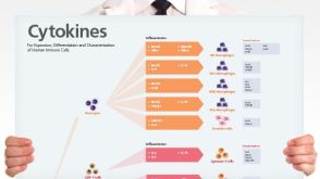
挂图
Human Immune Cytokines
Infographic of key cytokines for expansion, differentiation and characterization of major immune cell types
Megjugorac NJ et al. (MAY 2010)
Blood 115 21 4185--90
IL-4 enhances IFN-lambda1 (IL-29) production by plasmacytoid DCs via monocyte secretion of IL-1Ra.
The type-III interferon (IFN) family is composed of 3 molecules in humans: IFN-lambda1 (interleukin-29 [IL-29]),IFN-lambda2 (IL-28A),and IFN-lambda3 (IL-28B),each of which signals through the same receptor complex. Plasmacytoid dendritic cells (pDCs) are major IFN-lambda producers among peripheral lymphocytes. Recently,it has been shown that IFN-lambda1 exerts a powerful inhibitory effect over the T-helper 2 (Th2) response by antagonizing the effect of IL-4 on CD4(+) T cells and inhibiting the production of Th2-associated cytokines. Here,we asked whether Th2 cytokines exert reciprocal control over IFN-lambda production. IL-4 treatment during stimulation of human peripheral lymphocytes significantly elevated IFN-lambda1 transcription and secretion. However,pDCs were not directly responsive to IL-4. Using depletion and reconstitution experiments,we showed that IL-4-responsive monocytes are an intermediary cell,responding to IL-4 by elevating their secretion of IL-1 receptor antagonist (IL-Ra); this IL-1Ra acts on pDCs to elevate their IFN-lambda1 output. Thus,our experiments revealed a novel mechanism for regulation of both IFN-lambda1 production and pDC function,and suggests an expanded immunomodulatory role for Th2-associated cytokines.
View Publication
Pereira RC et al. ( 2016)
Frontiers in immunology 7 415
Human Articular Chondrocytes Regulate Immune Response by Affecting Directly T Cell Proliferation and Indirectly Inhibiting Monocyte Differentiation to Professional Antigen-Presenting Cells.
Autologous chondrocyte implantation is the current gold standard cell therapy for cartilage lesions. However,in some instances,the heavily compromised health of the patient can either impair or limit the recovery of the autologous chondrocytes and a satisfactory outcome of the implant. Allogeneic human articular chondrocytes (hAC) could be a good alternative,but the possible immunological incompatibility between recipient and hAC donor should be considered. Herein,we report that allogeneic hAC inhibited T lymphocyte response to antigen-dependent and -independent proliferative stimuli. This effect was maximal when T cells and hAC were in contact and it was not relieved by the addition of exogenous lymphocyte growth factor interleukin (IL)-2. More important,hAC impaired the differentiation of peripheral blood monocytes induced with granulocyte monocyte colony-stimulating factor and IL-4 (Mo) to professional antigen-presenting cells,such as dendritic cells (DC). Indeed,a marked inhibition of the onset of the CD1a expression and an ineffective downregulation of CD14 antigens was observed in Mo-hAC co-cultures. Furthermore,compared to immature or mature DC,Mo from Mo-hAC co-cultures did not trigger an efficacious allo-response. The prostaglandin (PG) E2 present in the Mo-hAC co-culture conditioned media is a putative candidate of the hAC-mediated inhibition of Mo maturation. Altogether,these findings indicate that allogeneic hAC inhibit,rather than trigger,immune response and strongly suggest that an efficient chondrocyte implantation could be possible also in an allogeneic setting.
View Publication

 EasySep™小鼠TIL(CD45)正选试剂盒


 挂图Human Immune Cytokines Infographic of key cytokines for expansion, differentiation and characterization of major immune cell types

 沪公网安备31010102008431号